Download
1 / 25
250 likes | 251 Views
Sunrise Free Radical School. Inflammation and Immunity: From Innate Oysters to Adaptive Humans. J. John Cohen Department of Immunology University of Colorado Medical School john.cohen@uchsc.edu. Metchnikoff 1883.

E N D
Sunrise Free Radical School Inflammation and Immunity: From Innate Oysters to Adaptive Humans J. John Cohen Department of Immunology University of Colorado Medical School john.cohen@uchsc.edu Inflammation and ImmunitySFRBM EducationProgram Cohen 1
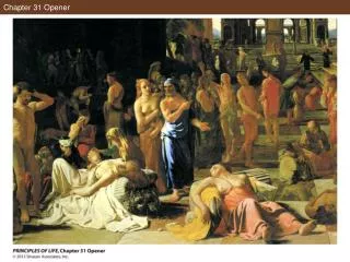
Metchnikoff 1883 Metchnikoff watched the reaction to a thorn inserted into a mollusk. Hemocytes (amoebocytes) arrived and tried to ingest the foreign body; if they could not, they walled it off, gradually converting into, or recruiting, fibroblasts. We do exactly the same thing to foreign bodies. Source: Wikipedia Inflammation and ImmunitySFRBM EducationProgram Cohen 2
Mouse macrophage Oyster hemocyte The similarity between an invertebrate phagocyte and our own is striking; they also use many of the same mechanisms, including the production of reactive oxygen species. http://www.mdsg.umd.edu/oysters/oysblood.htm http://itgmv1.fzk.de/www/itg/diabate/images.html#fig5 Inflammation and ImmunitySFRBM EducationProgram Cohen 3
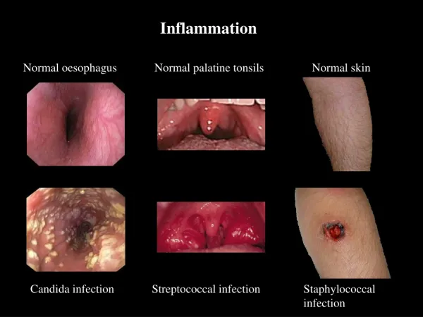
silicone droplets chromic catgut The clear (unstained) globules of silicone have been released from a ruptured breast implant. They also cannot be ingested, and this early stage shows the accumulation of macrophages, including one that has become a multinucleated giant cell showing the “asteroid bodies” characteristic of the foreign body reaction. Foreign body reactions in the human. The suture material is not digestible by macrophages, so it has been walled off by fibroblasts. Atlas of Granulomatous DiseasesYale Rosen, M.D. Inflammation and ImmunitySFRBM EducationProgram Cohen 4
Toll Like Receptors • The next slide shows a schematic that gives a feel for the multiple Toll-like receptor (TLR) pathways. • These receptors respond to foreign molecular patterns (PAMPs), so they are referred to as PRRs. • Recognized motifs include: • lipopolysaccharide (LPS) from Gram-negative cell walls, • peptidoglycans from the cell walls of both Gram-negative and – positive bacteria, • viral double-stranded RNA, and • CpG-rich bacterial DNA. • The result of the signals in all cases is a pro-inflammatory response and the release of chemokines and cytokines. • Review: Beutler B. Inferences, questions and possibilities in Toll-like receptor signaling. Nature. 2004 Jul 8;430(6996):257-63. Inflammation and ImmunitySFRBM EducationProgram Cohen 5
Ligands are PAMPs (pathogen- associated molecular patterns); Receptors are PRRs (pattern- recognition receptors) Inflammation and ImmunitySFRBM EducationProgram Cohen 6
The structure of two chemokines, HCC2 and, in the inset, eotaxin, shows their overall similarity. Their receptors are 7-span transmembrane structures which are, in general, ion channels that result in cell activation. Chemokines are low molecular weight peptides whose major role is in inflammation. CCR5, a chemokine receptor HCC2, a chemokine Source: Aegis (CDC) Source: Protein data base (PDB) Inflammation and ImmunitySFRBM EducationProgram Cohen 7
Human family Oyster family http://faculty.haas.berkeley.edu/arose/Asher1st2.htm In ecology, there are r-strategists and K-strategists. Oysters are r-strategists: they have huge numbers of progeny but invest relatively little in the survival of any individual; their survival is statistical. So they have the simple innate immune response, but cannot amplify it, nor do they adapt to specific challenges. The human is a K-strategist; small litters, but a heavy investment in individual survival. So we (jawed vertebrates) have added the adaptive immune response which vastly increases our chances (remember the Bubble Boy, born without an adaptive immune response and unable to live in the real world). Inflammation and ImmunitySFRBM EducationProgram Cohen 8
Dendritic cell iccosomes Dendritic cells are the link between innate and adaptive immunity. They are superb phagocytes. David Hunt. Cell Systems Initiative, U. of Washington. Inflammation and ImmunitySFRBM EducationProgram Cohen 9
Dendritic Cells • When dendritic cells are bathed in the cytokine and chemokine products of the innate response, they change, and move from the local area through the lymphatics to the draining lymph node (next slide) as they mature into the best antigen-presenting cells. • Iccosomes are clumps of stored antigen-antibody immune complexes, which allow the dendritic cell to stimulate immunity for a long time. • Dendritic cells enter the lymph node via the afferent lymphatics and percolate through the substance of the node, positioning themselves at the interface between T and B cell areas. There they display their processed antigenic peptides on both MHC Class I and Class II, so that it can be recognized by the best-fitting receptors of both CD4 (helper) T cells (which see antigen on Class II) and CD8 (killer) T cells (which see antigen on Class I). Inflammation and ImmunitySFRBM EducationProgram Cohen 10
chemokines cytokines PAMP PRR T Immature dendritic cell Mature dendritic cell Inflammation and ImmunitySFRBM EducationProgram Cohen 11
Antigen is picked up by a dendritic cell and taken to the draining lymph node, where it is presented to T cells. Artist: Helen Macfarlane Inflammation and ImmunitySFRBM EducationProgram Cohen 12
The peptides are now on the cell surface, presented on MHC Class II molecules and cross-presented on MHC Class I Inflammation and ImmunitySFRBM EducationProgram Cohen 13
LYMPH NODE A: Germinal centres (B cells) B: Zone of T, B, and DC contact C: Paracortex (T cells) D: Afferent lymphatics E: Subcapsular sinus F: Efferent lymphatic F Inflammation and ImmunitySFRBM EducationProgram Cohen 14
Th Th Th A helper T cell with the correct receptor recognizes peptide + MHC and becomes activated
T Cell Receptor MHC foreign peptide Dendritic Cell T Cell Inflammation and ImmunitySFRBM EducationProgram Cohen 16 Pique, Garcia, Wilson: Scripps
Members of the helper cell family • There are 3 cells in the helper family. • Th1 cells are the activators of delayed hypersensitivity and are sometimes thought of as “pro-inflammatory”. Their characteristic cytokine, interferon-gamma, is strongly chemotactic for macrophages. • Th2 cells help B cells make antibody. Their cytokines oppose Th1 development (Th1 cytokines oppose Th2 development, so there is “sibling rivalry”). Thus Th2 are sometimes thought of as anti-inflammatory. • Newly recognized are the Tregs, whose cytokines shut down both Th1 and Th2 responses. This is one of the hot areas in immunology. Inflammation and ImmunitySFRBM EducationProgram Cohen 17
Inflammation, delayed hypersensitivity APC Th1 IL-2, IFN-γ Downregulation of immunity APC IL-10, TGFβ Treg B cell help; sometimes suppression of inflammation IL-4, IL-5, IL-10 APC Th2 Inflammation and ImmunitySFRBM EducationProgram Cohen 18
T T T T T T T Activated T cells divide Inflammation and ImmunitySFRBM EducationProgram Cohen 19
T Activated Th1 cells release lymphokines Many macrophages are attracted Inflammation and ImmunitySFRBM EducationProgram Cohen 20
V V V All cells showpeptides from the proteins they make, including virus and tumor proteins, on MHC Class I molecules Inflammation and ImmunitySFRBM EducationProgram Cohen 21
CTL V V V Foreign or abnormal peptides + MHC are recognized by a cytotoxic (killer) T cell Killer T cells (cytotoxic T lymphocytes, CTL) actually don’t kill; they induce their targets to kill themselves by apoptosis. There are two pathways, one dependent on the exocytosis of granules, the other on transmembrane signaling via the Fas- Fas ligand interaction. Inflammation and ImmunitySFRBM EducationProgram Cohen 22
CTL V V V The CTL transmits a death signal Granule exocytosis Fas: Fas ligand Inflammation and ImmunitySFRBM EducationProgram Cohen 23
The abnormal cell dies by the process of apoptosis At the meeting a video of a CTL inducing apoptosis in a virus-infected cell was shown. It was obtained from Cells Alive! http://www.cellsalive.com/ctl.htm Inflammation and ImmunitySFRBM EducationProgram Cohen 24
Why adaptive? Amplification of inflammation T cell clones expand 64,000 x in 4 days Each activated Th1 cell attracts 1,000 macrophages Cytokine-stimulated macrophages are “angry” Repertoire Almost unlimited number of epitopes recognized Memory Secondary responses: sooner, steeper, faster, higher lower threshold Inflammation and ImmunitySFRBM EducationProgram Cohen 25