Download
1 / 22
240 likes | 669 Views
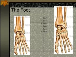
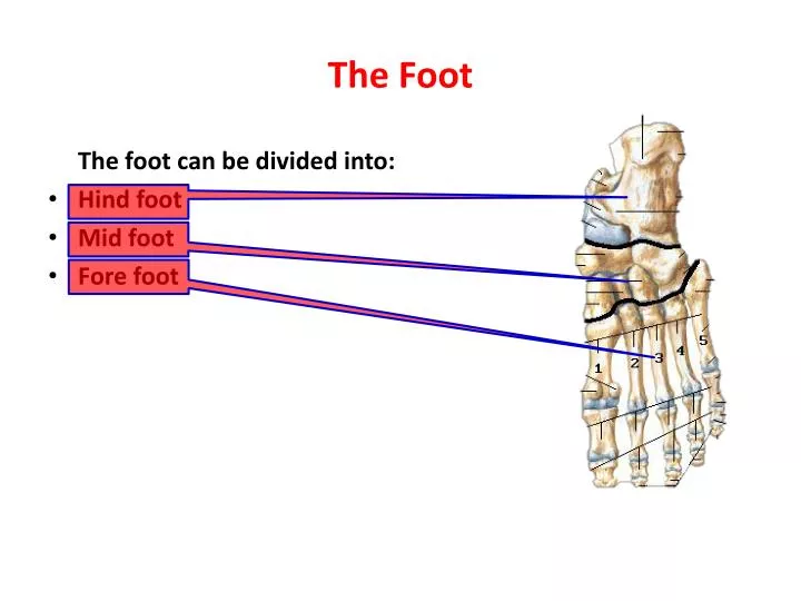
The Foot. The foot can be divided into: Hind foot Mid foot Fore foot. The Fascia of the Foot. The foot is covered by: Skin and superficial fascia Deep fascia It is continuous proximally with the extensor retinaculum

E N D
The Foot The foot can be divided into: • Hind foot • Mid foot • Fore foot
The Fascia of the Foot • The foot is covered by: • Skin and superficial fascia • Deep fascia It is continuous proximally with the extensor retinaculum It is also continuous laterally and posteriorly with the plantar fascia It is continuous distally with fibrous digital sheath which is interconnected with transverse metatarsl ligaments
Compartments of the Foot Vertical fibrous septa dividing the foot into: • Medial compartment • Central compartment • Lateral compartment In addition, • Interosseous compartment • Dorsal compartment
Medial compartment: Abductor hallucis, Flexor hallucisbrevis, tendon of flexor hallucislongus, medial plantar nerve and vessels • Central compartment: Flexor digitorumbrevis, tendons of flexor hallucislongus and flexor digitorumlongus, quadratusplantae, and lumrical muscles • Lateral compartment: Abductor and flexor digitiminimibrevis • Interosseous compartment: Dorsal and plantar interossei muscles, and deep plantar and metatarsal vessels • Dorsal compartment: Extensor hallucisbrevis and extensor digitorumbrevis and neurovascular structures of the dorsum of foot.
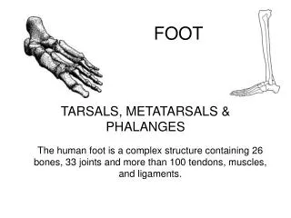
Muscles of the Foot • There are 20 muscles in the foot 14 plantar muscles arranged in four layers 2 dorsal muscles 4 foot intermediate in position • The muscles maintain the arches of foot • Support phase of stance • Weight received at the heel transferred to ball of foot and big toe • Adductor hallucis active during push off phase of stance in pulling 4 metatarsals towards the great toe fixing transverse arch of the foot and resists spread of metatarsal heads as force applied
First Layer Include the following 1. Abductor hallucis 2. Flexor digitorumbrevis 3. Abductor digitiminimi • The first two muscles are supplied by the medial plantar nerve, and the last one supplied by lateral plantar nerve • The action as the name implies 3 1 2
Second Layer • Quadratus Plantae helps in flextion of lateral four digits • Lumrical muscles flex the proximal digits and tetend other digits • They are supplied by lateral plantar nerve except the first lumrical muscle supplied by medial plantar nerve 2 1
Third Layer 1. Flexor hallucisbrevis 2. Adductor hallucis 3. Flexor digitiminimibrevis The first one and oblique head of the second muscle are supplied by medial plantar nerve, while the transverse head of the second muscle and the last one are supplied by lateral plantar nerve. 1 3 2
Fourth Layer • Plantar interossei • Adduct 3-5 digits • Dorsal interossei 4 muscles • Abduct 2-4 digits • All are supplied by lateral plantar nerve
Extensor hallucisbrevis • Arises from the calcaneum • Inserted to the big toe Extensor digitorumbrevis • Arises from the calcaneum • Inserted with long extensor to lateral four digits • Both are supplied by deep fibular nerve
Nerves of the Foot Cutaneous nerves: • Medially: saphenous nerve to the head of 1st metatarsal • Dorsally: superficial and deep fibular nerves • Inferiorly: medial and lateral plantar nerves • Laterally: Sural nerve • Posteriorly: medial and lateral calcaneal branches of tibial and sural nerves, respectively.
Ankle Joint • It is a synovial type of hinge variety • It is called (Talocrural Articulation) • The parts forming the joint are: Lower end of tibia and fibula Head of talaus Inferior transverse part of tibiofibularligament
The Capsule and Synovial Membrane • It is a sheath of dense connective tissue enveloping the knee joint • It is thin anteriorly and posteriorly • It is supported by medial and lateral collateral ligaments • Superiorly it is attached to superior borders of articular surfaces of tibia and malleoli • Inferiorly it is attached to the talus • The synovial membrane lines the capsule from all sites
Ligaments of Ankle Joint Lateral ligament consists of: 1. Anterior talofibular ligament It is flat and weak 2. Posterior talofibular ligament It is thick and strong It runs horizontally, medially then posteriorly 3. Calcaneofibular ligament 1 2 3
Ligaments of Ankle Joint Cont., • Medial ligament (Deltoid) • It is large and strong • It consists of: 1. Tibionavicular 2. Tibiocalcaneal 3. Anterior tibiotalar 4. Posterior tibiotalar • It stabilizes the ankle during eversion and prevent subluxation (partial dislocation) 4 3 1 2
Movements of the ankle Joint • Dorsiflexion • Plantarflextion • Eversion • Inversion
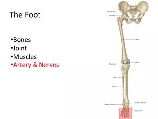
Neurovascular Supply • Blood supply: Malleolar branches of fibular and anterior and posterior tibial arteries • Nerve supply: Tibial and deep fibular nerves
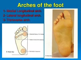
Major Ligaments of the Foot 1. Plantar calcaneonavicular ligament (Spring Ligament) 2. Long plantar ligament 3. Plantar calcaneocuboidal ligament (Short plantar Ligament) 3 2 1
Arches of the Foot Longitudinal arches Medial longitudinal arch • It is higher and more important • Talar head is the key stone of the arch • It is made of calacaneum, talaus, navicularbone, the three cuneiform bones, and medial three metatarsal bones • Tibialis anterior attached to 1st meta tarsal and medial cuneiform strengthen the arch • Fibularislongus crossing from lateral to medial side support the arch