Download
1 / 58
1.88k likes | 4.77k Views
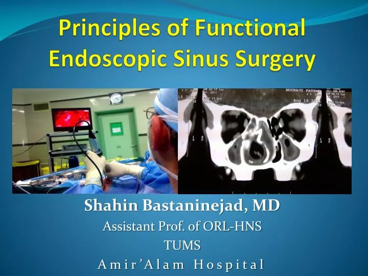
Principles of Functional Endoscopic Sinus Surgery. Shahin Bastaninejad , MD Assistant Prof. of ORL-HNS TUMS Amir’Alam Hospital. Outline. Definition Anatomy Patient evaluation FESS Concepts of Surgery. Definition. Functional Endoscopic Sinus Surgery.

E N D
Principles of Functional Endoscopic Sinus Surgery ShahinBastaninejad, MD Assistant Prof. of ORL-HNS TUMS Amir’Alam Hospital
Outline • Definition • Anatomy • Patient evaluation • FESS Concepts of Surgery
Functional Endoscopic Sinus Surgery • Replaced old practice of obliterating sinuses and removing mucosa. Concept of irreversibly diseased mucosa refuted. • Functional aspect refers to: • Preserving normal structures • Removing only obstruction • Preserving mucosa • Attempt to restore function
Ethmoid anatomy is complex: Labyrinth Lamellae 1st - Uncinate 2nd - Ethmoid bulla 3rd - Basal lamella of middle turbinate 4th - Superior turbinate Ethmoid anatomy
Drainage • Frontal, anterior ethmoid & maxillary – OMC • Posterior Ethmoids – Superior meatus • Sphenoid sinus – Sphenoid-ethmoidal recess
Middle Turbinate • Three components • First – Anterior, oriented in a sagittal plane and attached to skull base • Second – Middle, oriented in a Vertical plane and attached to lamina papyracea (basal lamella and separates ant from post ethmoids) • Third – Posterior, oriented in a horizontal plane and attaches to perpendicular plate of palate (forms roof of middle meatus, anterior to sphenopalatine foramen)
Ostiomeatal Complex (OMC) • Common drainage for frontal, maxillary and anterior ethmoid sinuses.
OMC • Infundibulum – funnel shaped area whereby the maxillary, ant ethmoid and frontal sinuses drains • Uncinate process– Sickle shaped bony ethmoidal structure • Hiatus Semilunaris– Half-moon shape opening of infundibulum
Uncinate Process • Attaches to the following structures: • Inf & far post. – To ethmoid process of inf. Turb
Uncinate Process • Ant & far sup. – To lamina papyracea, skull base or mid turb
Uncinate Process 52%
Bulla Ethmoidalis • The greatest anterior ethmoid air cell, attached to lamina papyrcea and usually open into lateral sinus
Middle turbinate: Horizontal and vertical basal lamella SBR Sinus Lateralis RBR
Sphenoid Ostium • Medial to posterior sup. turbinate • Located between nasal septum and inferior aspect of sup. turbinate • Located at the same level as the roof of the maxillary sinus • Located 4 microdebrider/suction tip breaths above the choanae • Located 7cm from nasal crest at 30°
Sphenoid Sinus • Relationships of important structures: • Optic nerve – superior-lateral • Carotid artery/cav sinus – mid-lateral • Vidian nerve and maxillary nerve – inferior-lateral
Square – ant clinoid process, Circles – optic canals, triangle – vidian nerve Asterisk – pneumatization of pterygoid process
Presellar Sellar Conchal Post sellar
Cribriform plate Keros classification 1-3mm 3-7mm 7-16mm
Keros Classification • Type I • 1-3mm • Type II • 3-7mm • Type III • 7-16mm
Kuhn Cells Frontal Cells
Frontal Recess • Anatomic Boundries: • Ant – unicate process & aggernasi • Post – bulla ethmoidalis and suprabullar lamella • Lateral – lamina papyracea • Medially – hiatus semilunaris or middle turb • Inf – Ethmoidinfundibulum • Sup – Fovea ethmoidalis, supraorbital air cell, anterior ethmoid artery and frontal ostium
Draf IIA Draf I
Draf III Draf
Frontal Sinus – Mucociliary Pattern Save Mucosal Layer in Lateral part while performing Draf III opertation
Pre-op CT Evaluation • CLOSE Technique • C – Cribriform • L – Lamina Papyracea • O – Orbits, onodi cell, Optic Nerve • S – Sphenoid, Skull Base • E – Ethmoid Arteries
C - Cribriform • Assess the Keros type • Look for assymetry
L – Lamina Papyracea • Check for dehiscence or pathologic fractures
O – Orbit, Optic Nerve, Onodi Cells • Check for dehiscence • Assess for onodi cells (superior-lateral to sphenoid) • Orbital slope
S – Sphenoid, Skull base • Assess for Carotid dehiscence and aeration patterns • Conchal, Pre-sellar, & Sellar (thickness of clivus)
Skull base • Assess slope of skull base • Assess if roof of sphenoid is level with skull base
FESS Concepts of surgery