Download
1 / 33
330 likes | 399 Views
Biomedical Innovations Unit 3 Tissues of Life A Focus on Blood. Blood Introduction. Blood is a special C onnective T issue , and is the major component of the Circulatory System

E N D
Biomedical Innovations Unit 3Tissues of LifeA Focus on Blood
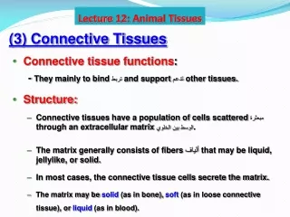
Blood Introduction Blood is a special ConnectiveTissue, and is the major component of the Circulatory System Connective tissue is a group of cells that collectively function to support, connect, and/or separate other tissues and organs. The Circulatory System is comprised of two sub-systems: 1. Cardiovascular System Includes network of blood vessels, blood, and heart Major function is to transport nutrients, gases and hormones to cells and wastes from cells for excretion outside the body 2. Lymphatic System Includes network of lymph vessels, the lymphocyte white blood cell, and lymphoid organs(tonsils, spleen, thymus, bone marrow, and lymph nodes) Major functions are to return fluid that escapes from blood vessels back to the bloodstream AND fight infections and give immunity to disease
Functions Of Blood 1. Transportation Blood transports dissolved gases, nutrients, hormones and metabolic wastes 2. Protection & Clotting White Blood Cells (WBC) protect the body against foreign molecules Platelets (cell) and clotting proteins in blood minimize blood loss when a blood vessel is damaged (clot) 3. Regulation Blood regulates the pH and electrolyte composition of the interstitial fluids (fluid between cells) Blood regulates body temperature: transfers heat via counter-current exchange
Composition of Blood Contains cellular and liquid components Liquid Portion: ~ 55% plasma Cellular Portion: ~ 45% formed elements Normal blood pH is ~7.35-7.45 (neutral) Blood volume Varies inversely with body fat Blood volume as body fat Males typically have 5 to 6 liters (~10.5 to 12.5 pints) Females typically have 4 to 5 liters (~8.5 to 10.5 pints) How can blood volume be determined? How much is a “unit” of donated blood? • Volume: • Assess: Blood pressure • Calculate: Radioactive dye • Units: • 1 unit donated = ~ 1 pint (0.5L) • 1 unit accepted = ~0.75 pint • Packed RBC (prBC)
Composition of Blood • 55% Plasma • 92% - Water • 7% - Proteins (fibrinogen, hormones, albumins & globulins) • 1% - other solutes (ions, gases, nutrients, wastes, etc.) • 45% Formed Elements • 99.9% - erythrocytes (Red Blood Cells - RBCs) • 0.1% - leukocytes (White Blood Cells - WBCs) & thrombocytes (Platelets)
Composition of Blood - Plasma Figure 19.1b
Composition of Blood – Formed Elements Figure 19.1c
ID the Formed Elements Be able to identify any of the formed elements to RBC, WBC, or Platelet.
Lecture 1a - Review Break • Visualize the Composition of Blood • Microscopy & Blood Cell Identification Lab
Lecture 1b – Overview: Composition of Blood Hematocrit or Packed Cell Volume (PCV) measure of % RBC Males: 47% ± 5% Females: 42% ± 5% Figure 17.1
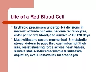
Erythrocytes – Red Blood Cells (RBCs) Oxygen-transporting cells 7.5µm in diameter (diameter of capillary 8 – 10µm) Most numerous of the formed elements Females: 4.3 – 5.2 million cells/mm3 Males: 5.2 – 5.8 million cells/mm3 Made in the red bone marrow in long bones, cranial bones, ribs, sternum, and vertebrae Average lifespan is 100 – 120 days
RBC Structure And Function Have no organelles or nuclei Significance? True for all species? Hemoglobin – oxygen carrying protein Each RBC has 200-300 million hemoglobin molecules Biconcave shape Significance?
Hemoglobin • Comprised of four protein chains, each called a globin. • Each globin is bound to a red pigment, called a heme molecule. • Contains a single Fe atom • Each Fe atom can bind to a single O2 molecule • How many O2 molecules can each hemoglobin combine with? • What is the term for when hemoglobin binds with O2? • CO2? • Are either a reversible reaction?
Leukocytes – White Blood Cells (WBCs) Protect the body from: infectious microorganisms Cancerous cells Foreign particles Typically, function outside the bloodstream in loose connective tissue Diapedesis - circulating leukocytes leave the capillaries and enter the interstitial fluid Exception? WBCs have a nucleus and are larger than RBCs Most produced in bone marrow Exception? Lifespan of 12 hours to several years
Leukocytes – White Blood Cells (WBCs) Two types of leukocytes Granulocytes Agranulocytes Relative WBC Count Never Let Monkeys Eat Bananas Figure 17.5
Lymphocyte Compose 20 – 45% of WBCs The most important cells of the immune system Nucleus – stains dark purple Effective in fighting infectious organisms Act against a specific foreign molecule (antigen) Two main classes of lymphocyte T cells – attack foreign cells directly Active in cell mediated immune response B cells – multiply to become plasma cells that secrete antibodies Active in the humoral immune response Figure 17.4d
Platelets Structure Small, nearly colorless bodies appearing as irregular spindles or oval disks (~2-4 μm) originate in bone marrow from giant cell megakaryocyte Functions Hemostasis Regulation of blood flow Coagulation, or blood clotting
Summary of Formed Elements Table 17.1
Review Activity Break • Blood Disorders
Blood Cell Formation Hematopoiesis – process by which blood cells are formed 100 billion new blood cells formed each day Takes place in the red bone marrow of the humerus, femur, sternum, ribs, vertebra and pelvis Red marrow – actively generates new blood cells Contains immature erythrocytes Remains in epiphyses, girdles, and axial skeleton Yellow marrow – dormant (can become active if needed) Contains many fat cells Located in the long bones of adults
Cell Lines in Blood Cell Formation All blood cells originate in bone marrow All originate from one cell type Blood stem cell (pluripotential hematopoeitic stem cell) Lymphoid stem cells - give rise to lymphocytes Myeloid stem cells - give rise to all other blood cells
Cell Lines in Blood Cell Formation Genesis of erythrocytes (erythropoiesis) Committed cells are proerythroblasts Remain in the reticulocyte stage for 1–2 days in circulation Loss of nucleus Formation of leukocytes (leukopoiesis) Granulocytes form from myeloblasts Monoblasts enlarge and form monocytes Platelet formation (thrombopoiesis) Form from megakaryoblasts break apart into platelets
The Blood Throughout Life First blood cells develop with the earliest blood vessels Late in the second month the liver and spleen take over blood formation Bone marrow becomes major hematopoietic organ at month 7
RBC life span and circulation Replaced at a rate of approximately 3 million new blood cells entering the circulation per second Damaged or dead RBCs are recycled by phagocytes Components of hemoglobin individually recycled Heme stripped of iron and converted to biliverdin, then bilirubin Iron is recycled by being stored in phagocytes, or transported throughout the blood stream bound to transferrin
Red Blood Cell Turnover Figure 19.5
Clotting Mechanisms • Know the general stages of blood clotting • Stage 1: Source of damage • Stage 2: prothrombin thrombin • Calcium, prothrombin activator • Stage 3: fibrinogen fibrin • Calcium, thrombin • Be able to identify the key difference between intrinsic and extrinsic pathways • Stage 1
Clotting Cont. • What two conditions increase clotting? • What two conditions decrease clotting? • How are clots removed? • Fibrinolysis
You should be able to… Identify and describe the following blood disorders/conditions: Identify and describe: The different types of tissues Functions of the blood Blood composition Plasma & Formed Elements % Hematocrit Blood Cell Formation The process of clotting Blood type based on tests and genetic inheritance Antigen vs. antibody Coagulation vs. agglutination • Leukemia • Leukopenia • Leukocytosis • Anemia • Polycythemia • Blood doping • Sickle-cell anemia • Embolus • Thrombus • Erythroblastosis fetalis