Download
1 / 26
260 likes | 809 Views
Assessment of the Cardiovascular System. Health Assessment NU 325 SU 2010. Assessing the Pattern – Activity & Exercise (Review). Should start at the initial interaction with the client (interview/health history to PE) Goals of assessing the pattern

E N D
Assessment of the Cardiovascular System Health Assessment NU 325 SU 2010
Assessing the Pattern – Activity & Exercise(Review) • Should start at the initial interaction with the client (interview/health history to PE) • Goals of assessing the pattern • What data collection methods or sources of data you will utilize in assessing the pattern, e.g. cardiovascular function? • Subjective and objective data (including activity/exercise tolerance) • Risk factors • Familial, environmental, behavioral, psychological, etc
Physical Examination of the Cardiovascular System: Learning Objectives • Describe the normal physical characteristics of the heart and precordium • Describe the physiologic events that generate normal and abnormal heart sounds and murmurs • Demonstrate the process of physical examination of the heart and precordium • Describe abnormalities detected by precordial assessment • Describe how to measure central venous pressure by examination of the neck veins
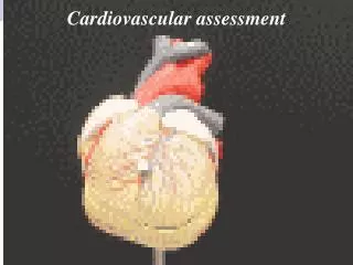
Overview of the Anatomy & Physiology of the Cardiovascular System • The heart and great vessels • Blood vessels: arterial and venous • Blood flow • The conduction system and the cardiac cycle Base Apex
Two phases (“lub-dub” on auscultation: normal heart sounds) • Ventricular systole (S1: “lub”) – closing of MV and TV • Ventricular diastole (S2: “dub”) – closing of the AV and PV • Abnormal heart sounds: • Murmurs • Extra heart sounds: S3, and S4
Murmurs • Altered Blood Flow • (Murmurs) • Origins of murmurs: • High flow rates • Forward flow • Backward or • regurgitant
General Principles of Assessment of the Cardiovascular System • Precordial examination • Focusing on the anterior chest wall and determine the status of underlying cardiovascular structures • PE Technique: Inspection, Palpation, Auscultation • Arterial pulse examination • Skin is inspected during cardiovascular examination • Neck vein (venous pulse) examination • Determine the characteristic of venous pulsations and central venous pressure (CVP) • Integrate this when assessing person’s neck
General Principles of Assessment of the Cardiovascular System • Equipment • Stethoscope with bell and diaphragm • Ruler • Light source for tangential lighting • Doppler (optional) • Patient preparation • Quiet room, privacy, positioning: sitting or lying on exam table or bed • Adequate lighting and exposure of body parts • Precordial examination position: examiner stands on right side • Precordial landmarks
Aortic area Pulmonic area Tricuspid area Mitral area PMI Sternoclaviculular area Right ventricular area Erb’s point p. 302-303 Precordial Landmarks
General Principles of Assessment of the Cardiovascular System • Examination and documentation focus • Precordium • Inspection: pulsatile movements • Palpation: pulsatile movements, vibrations • Auscultation: heart rate and rhythm, heart sounds and murmurs • Arterial pulses (previously discussed) • Neck veins • Pulse contour and amplitude • Distention • Height of venous pulsation Note: Palpate and auscultate carotid arteries (previously discussed)
Assessment of the Heart and Precordium PROCEDURE
Procedure 1. Inspect the precordium • Normal findings: • Location of apical pulse or PMI (5th ICS, MCL) • Pregnancy, obese, large breasts • Deviations from normal: • Displaced PMI • Heave or lift (right or left) 2. Palpate major precordial landmarks • Aortic, pulmonic, Erb’s point, tricuspid, mitral (use ball of your hand) • Note any pulsations, thrills, or rubs: describe location, amplitude, duration, and direction of impulse • Perform palpation in 3 different positions: supine, forward sitting or left lateral decubitus
Procedure Findings on palpation of precordial landmarks: • Normal findings: • Pulsatile movements (PMI area) • Deviations from normal: • Vibrations or palpable thrills 3. Auscultate the major precordial landmarks • Sequence: aortic, pulmonic, Erb’s point, tricuspid, mitral • Listen several cardiac cycles at each landmark (one sound at a time) • Use diaphragm of stethoscope to detect higher-pitched sound (S1, S2) • Use bell of stethoscope to detect lower-pitched sound (S3, S4) • Clinical significance: eliciting heart rate and rhythm
4. Identify S1 and S2 Normal finding: S1, loudest at the apex (mitral area) S2, loudest at the base (pulmonic area) Note intensity and splitting (physiologic split) Note abnormal heart sounds Abnormal findings Splitting (S1 or S2) Third heart sound (S3) Fourth heart sound (S4) Opening Snap Summation gallop Ejection click Midsystolic click Pericardial friction rubs Murmurs Procedure
Abnormal Heart Sounds Third heart sound (S3) • Also called, ventricular gallop, a low-frequency sound best heard using the bell of the stethoscope at either the left or lower right apical area • Sound may be accentuated during inspiration (sounds like “Ken-tuc-ky”) • Normal in young children or in people with high cardiac output • Clinical significance: CHF, tricuspid or mitral insufficiency • The early diastolic represents rapid ventricular filling, vibrations caused by blood forcefully hitting the ventricular wall
Abnormal Heart Sounds Fourth heart sound (S4) • Also known as atrial gallop, occurs near the end of diastole when the atria contract • A low frequency sound (use bell of stethoscope) • LV S4loudest at the apical area: supine or left lateral decubitus position • RV S4 loudest at the lower right ventricular area when person assumes a supine position • May increase volume during inspiration (sounds like “Ten-nes-see”) • Clinical significance: hypertension, aortic and pulmonic stenosis, acute myocardial infarction • S4 occurs after atrial contraction, caused by vibrations when blood flows rapidly into the ventricles • Vibrations result from the flow of a high blood volume or if ventricle wall has low compliance
Abnormal Heart Sounds Murmurs • Timing – which phase of the cardiac cycle? • Intensity – how loud is the murmur? Use murmur grading system I – barely audible II – audible but quiet III – clearly heard IV – Loud; may be associated with thrill V – Very loud; palpable thrill; may hear with stethoscope partially off chest VI – Very loud; palpable thrill; can hear without using a stethoscope Documentation example: (+) S3 A2 murmur III/VI 2nd ICS RPSB (right parasternal border)
Abnormal Heart Sounds Murmurs • Quality – what is the quality, pitch, and pattern of the murmur? • Describe pitch as high or low, quality as blowing, harsh, or musical • Patterns to changes in the murmur intensity, e.g. crescendo, decrescendo • Location – over which precordial landmark is the murmur loudest? • Radiation –is the sound of the murmur transmitted to other areas of precordium? • Ventilation – is the murmur affected by inspiration, expiration or position changes?
Abnormal Heart Sounds Common Types of Murmurs • Physiologic murmurs (detected during systole) – or known as non-pathologic or innocent murmurs, e.g. pregnancy, children, increased cardiac output states, etc. • Mitral regurgitation – pansystolic (holosystolic) • Aortic stenosis – mid-systolic ejection murmurs • Aortic regurgitation – pandiastolic Nurse’s role: • Recognition of life-threatening murmurs such as in patients who has recently had a MI (e.g. mitral regurgitation)
Procedure 5. Identify normal splitting of the first and second heart sounds • Split second sound (physiologic splitting of S2) most common, specially in young children • S2 is made up of 2 components • Mechanism: • Inspiration, venous return increased (PV delayed closure, AV closes first) • Expiration, sound occurs as one • Ask patient to take a deep breath and hold
5. Identify extra heart sounds and murmurs (other than physiologic split) 6. Auscultate precordium with person assuming different position (optional) Forward sitting, brings base of the heart closer to chest wall Left-lateral decubitus position, brings the apex of the heart closer to chest wall Procedure
Assessment of Neck Veins: Procedure Observe jugular venous pressure (JVP), commonly know as JVD • Assess by inspection • Venous pulse is not normally visible with person sitting fully upright • Observe the person from the right side • Positioning: HOB 30 to 45 degrees head slightly to the left • Provide tangential lighting to neck area Abnormal findings: (+) increase in JVP;TV dysfunction, right-sided heart failure, pulmonary hypertention, cardiac tamponade, etc
Estimate central venous pressure (CVP) by measuring the height of pulsation in the internal jugular vein (IJ) Select a reference point (sternal angle = 5 cm above the right atrium) Measure the distance (in cm) from the sternal angle to the top of distended jugular vein Add 5 cm to the value obtained, this is a rough estimate of CVP CVP ( 3-5 cm above sternal angle) Abnormal CVP is associated with same conditions as elevated JVP Assessment of Neck Veins: Procedure p. 319
Assessment of Neck Veins: Procedure Check for hepatojugular reflux • Indicated if a right sided heart failure is suspected • Procedure: • HOB: 30 to 60 degrees • Compress right upper quadrant for 30 to 60 seconds with your palm • (+) hepatojugular reflux; if JVP rises with this maneuver
Nursing Diagnoses Related to Cardiovascular Assessment • Decreased Cardiac Output • Signs and symptoms? (Subjective and Objective) • Altered Tissue Perfusion: acute or chronic • Chronic altered: • cerebral perfusion • cardiopulmonary perfusion • GIT tissue perfusion • renal tissue perfusion • peripheral tissue perfusion related to n interruption of arterial flow and/or venous flow The above affects/impairs person’s functional pattern: activity/exercise
Homework • Readings • Documenting cardiovascular examination findings: F/S p. 319-320 • Read, and listen “Heart Sounds” posted by Professor Hollywood (scroll down under Course Documents) • Practice with Lab partner and SinMan for abnormal heart sounds