Download
1 / 43
440 likes | 686 Views
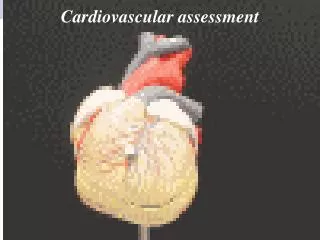
ASSESSMENT OF CARDIOVASCULAR FUNCTION. NUR240 Lecture 1. LECTURE OBJECTIVES. Review anatomy & physiology of the cardiovascular system. Discuss relevant aspects of the patient history. Describe physical assessment of cardiovascular status.

E N D
ASSESSMENT OF CARDIOVASCULAR FUNCTION NUR240 Lecture 1
LECTURE OBJECTIVES • Review anatomy & physiology of the cardiovascular system. • Discuss relevant aspects of the patient history. • Describe physical assessment of cardiovascular status. • Review diagnostic procedures, tests and medications relative to the cardiovascular system.
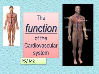
Anatomy & Physiology (What makes it “tick”!) Functions of the heart & CV system • Pumps blood to tissues to supply O2 & nutrients • Remove CO2 & metabolic wastes
Anatomy & Physiology CARDIAC CELLS HAVE UNIQUE PROPERTIES • AUTOMATICITY CELLS CONTRACT INDEPENDENTLY (THEY INITIATE THEIR OWN IMPULSE) • EXCITABILITY ION SHIFT • CONDUCTIVITY TRANSMIT IMPULSE TO ANOTHER CARDIAC CELL • CONTRACTTILITY HOW WELL THE CELL CONRACTS
Anatomy & Physiology PERICARDIUM / PERICARDIAL SAC • Protects heart from trauma • Serous fluid lubricates and prevents friction • Prevents heart from over filling
CORONARY ARTERIES Right & Left arteries encircle the heart and supply blood to the myocardium during ventricular relaxation( diastole) LEFT MAIN CORONARY ARTERY L ANTERIOR DESCENDING (LAD) L CIRCUMFLEX (LCX) RIGHT CORONARY ARTERY POSTERIOR MARGINAL
CORONARY ARTERIES (L) ARTERY CIRCUMFLEX (R) ARTERY LAD
CONTRACTION OF CARDIAC MUSCLE The heart can’t pump unless an electrical stimulus occurs Action Potential (AP) – electrical change (depolarization = contraction) Brought about by release of calcium (+ charge) into cells- mechanical change Intrinsic Pacemakers – depolarize and generate the AP
CONTRACTION OF CARDIAC MUSCLE The pacemaker with the fastest rate of depolarization stimulates the AP • SA node (60-100 bpm)- Upper R atrium- capable of initiating electrical impulse • AV node (40-60 bpm)- Lower R atrium • Other pacemakers ( 40 bpm) -what can affect SA/AV node function ?
DISRUPTION IN SERUM ELECTROLYES CAN RESULT IN ALTERATION IN CARDIAC CYCLE • Potassium • Calcium • Sodium • Magnesium
MONITORING MOVEMENT OF THE CRDIAC ACTION POTENTIAL (AP) • EKG – monitors the movement of the AP, in other words, the electrical changes. • How are the mechanical changes ( cardiac output ) monitored ?
CARDIAC CYCLE CARDIAC CYCLE – all the activities occurring in the heart during one contraction, and subsequent period of relaxation. Graphically represented on an EKG (ECG)
CARDIAC CYCLE EKG – A 12 lead EKG is a graphic record of the electrical forces produced by the heart
CARDIAC CYCLE Polarized (resting) cell – represented on EKG as baseline or isoelectric line Depolarization – impulse over specialized cardiac cells (not neuromuscular impulse) Repolarized cell – returns to normal. Na moves out of cell, K moves in – requires ATP How will ischemic tissue change the cardiac cycle ?
ELECTRODE POSITIONS “LEADS” • Leads measure electrical activity between 2 points • Movement toward electrode causes positive deflection • Movement away from electrode causes negative deflection
ELECTRODE POSITIONS A 12 Lead EKG shows electrical activity from 12 different positions in the heart, concentrating on (L) ventricle A 14 Lead EKG includes (R) ventricle activity
Cardiac output • SV- • CO- • Preload- • Afterload- • Ejection fraction • GOAL is to maintain adequate MAP so perfusion of oxygenated blood to vital organs occurs
Regulation of cardiac function & BP • Autonomic nervous system • Sympathetic norepinephrine • Parasympathetic – acetylcholine • Stimulation of adrenals by SNS – norepinephrine • Peripheral baro receptors • Stretch receptors • chemorecptors • hormones
STROKE VOLUME (SV) & CARDIAC OUTPUT (CO) • SV – amount of blood ejected by 1 ventricle in 1 beat • CO – volume ejected in 1 min Control of SV and HR = SV&HR are continually adjusted by the body, and are affected by the return of blood from the tissues (think of exercise) CO = SVxHR
STROKE VOLUME (SV) & CARDIAC OUTPUT (CO) Extrinsic control of HR is a more powerful way of controlling CO than changing SV • CVP causes stretching of (R) atrial muscle which stimulates SNS & HR (to help pump all the blood returned to it) • Remember “Starling’s Law”
STROKE VOLUME (SV) & CARDIAC OUTPUT (CO) • Stretch baroreceptors (aorta & carotid) detect in pressure which stimulates SNS & HR (to ensure adequate blood supply to heart/ brain) • If pressure detected, then PSNS is stimulated & HR is slowed (vagus nerve) (prevents excess arterial pressure which can damage organs)
CARDIAC LOAD Preload = degree of myocardial fiber stretch at the end of diastole and just before contraction Afterload = pressure against which ventricles must eject blood. This pressure is affected by systemic vascular resistance (SVR)
Age related changes • Decreased myocardial contractility • Thickening of endocardium & valves • Coronary arteries rigid & thickened • Decreased elasticity of vessel walls • Decreased internal diameter of vessels
CARDIAC ASSESSMENT Cardiac status of all patients should be routinely assessed. Everyone has a • Objective • Subjective CP Dyspnea Fatigue What else ?
IMMEDIATE NURSING INTERVENTIONS FOR ACUTE CARDIAC EVENTMOVIE Acronym M- Monitor for pain O- O2 and pulse ox V- Vital signs I- Intravenous fluids E- EKG monitoring Anything else??
Pain Assessment SLIDA or Precipitating/alleviating factors Quality Radiation Severity Timing
OTHER ELEMENTS OF CARDIAC ASSESSMENT • Previous cardiac hx • Other medical conditions that may affect heart function • Chest injury • Previous heart surgery • Past medical hx • Medications: prescribed, OTC, herbals • Activity tolerance • Health habits • Family hx
EXAMINATION • Inspection • Palpation • Percussion-? • Auscultation = S1, S2 at PMI Aortic Pulmonic Tricuspid Mitral
Heart RhythmRegular, Irregular, Regular Irregular Abnormal Sounds: Gallops Murmurs Bruits S3 ventricular gallop – heard in early diastole S4 atrial gallop – generally abnormal
Assessment of Murmurs Turbulent blood flow in valvular disorders and septal defects Timing of murmurs is a must! Systolic murmurs occur between S1 & S2 Diastolic murmurs occur between S2 & S1 Grade 1 – 6 identifies intensity of murmur
Other assessments • Jugular vein pressure – assess JVD which reflects increased filling volume and pressure on (R) side of heart JVD associated with (R) HF, SVC obstruction (Normal is 3-10cm H20) • Pulse deficit – the difference between apical HR and peripheral pulse-associated with Afib, and heart blocks • Pulse pressure – the difference between systolic & diastolic pressure
Other assessments • Respiratory: Lung sounds = rate, rhythm, quality, sputum • GI-Abdomen • Peripheral Vascular:Lower extremities
Diagnostic Procedures • EKG 12 Lead continuous cardiac monitoring holter monitor • Chest x-ray – detects enlargement of heart & pulmonary congestion
Diagnostic procedures • Echocardiography – ultrasound that reveals size, shape and motion of cardiac structures Evaluates heart wall thickness, valve structure, differentiates murmurs • TEE – transesophageal echocardiography provides a clearer image because less tissue for sound waves to pass through
Diagnostic procedures • Angiography / cardiac catherization determines coronary lesion size, location, evaluate (L) ventricular function, measures heart pressures • Exercise tolerance test • Radionuclide Imaging
Lab Studies Cardiac enzymes = enzymes are released when cells are damaged (MI). Enzymes are found in many tissues/muscles, and some are specific to cardiac tissue. Serial measurement can aid in dx, and monitor course of MI Cardiac enzymes = CPK – MB (CK-MB),myoglobin, Troponin In general, the greater the rise in the serum level of an enzyme, the greater the degree or extent of damage to the muscle. LDH
LAB studies • Electrolytes • Lipid panel • CBC • C – Reactive Protein • BNP- Human B-Natriuretic Peptide • Blood coags-PT/PTT/INR
NCLEX TIME Mary is attending a sophomore level nursing class on anatomy and physiology. Which statement, if made by Mary, demonstrates a good understanding of the anatomy and physiology of the heart? A."The heart is encapsulated by a protective coating called the endocardium.“ B."The SA node is considered the main regulator of heart rate.“ C."The left atrium receives deoxygenated venous blood from all peripheral tissues.“ D."Stroke volume is the amount of blood ejected by the right ventricle during each diastole
NCLEX TIME Kirsten is completing her graduate clinical rotation in a large urban teaching hospital in a medical coronary care unit (CCU). Which observation demonstrates a good understanding of completing a thorough cardiac examination? • A. In an obese client, an adult cuff size of 12 to 14 cm is preferable. • B.The carotid artery on the neck is auscultated to assess for the presence of a bruit. • C.The apical impulse is auscultated over the fifth intercostal space in the midclavicular line. • D.Palpation is used to determine cardiac size.
NCLEX TIME Edward is a 40-year-old white male. He is an accountant who works on average 11 hours per day. He reports feeling stressed each day, even with mundane things such as a traffic jam. His father had a massive myocardial infarction at the age of 48. His mother has a history of congestive heart failure. He seldom has time to exercise, but does eat balanced meals when possible, although he does not get to eat three meals a day. Select all factors that place Edward at risk for heart disease. • A.Family history • B.Age • C.Coping-stress tolerance • D.Race • E.Occupation