Download
1 / 28
290 likes | 444 Views
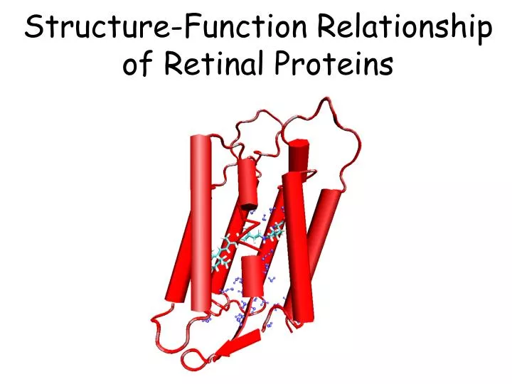
Structure-Function Relationship of Retinal Proteins. Structure of Retinal Proteins. C. F. B. A. E. D. G. GPCRs.

E N D
Structure of Retinal Proteins C F B A E D G GPCRs Retinal proteins or rhodopsins belong to the superfamily of seven-transmembrane helical (7TM) proteins. Seven helices, with N-terminus on the extracellular side and C-terminus on the cytoplasmic side of the membrane (not necessarily G-protein coupled)
Retinal Proteins -- Rhodopsins • Covalently linked to a lysine • Usually protonated Schiff base • all-trans and 11-cis isomers Chromophore
Bacteriorhodopsin -- bR • The simplest ion pump in biology • The simplest photosynthetic center • The best characterized membrane protein • Technological applications in molecular electronics • The first membrane protein with a known atomic-detail 3D structures
bR role in Bioenergetics Light Halobacterium Salinarum The Purple Membrane hn hn Cytoplasmic side [H+] H+ [H+] Extracellular side Proton Gradient ATP ADP ATP Synthase
Schematic proton path in bacteriorhodopsin H+ H+ Cytoplasmic side Transmambrane helices Transmambrane helices Extracellular side
Active Channels Need a ‘Switch’ Mechanism H+ H+ H+ hn H+ H+ H+ H+ H+ H+ H+ H+ H+ H+ H+ What is the switch in bR? How does it work?
Photocycle of bR Photo-induced 5 ms 3 ps 1 ms 5 ms 40 ms 5 ms All intermediates are trapped in low temperature and have been characterized by vibrational and absorption spectroscopy.
No membrane protein has been studied as extensively as bR Photo-induced 5 ms 3 ps 1 ms 5 ms 40 ms 5 ms All intermediates have also been characterized by X-ray crystallography!
Schematic proton path in bacteriorhodopsin Cytoplasmic side Transmambrane helices Transmambrane helices Extracellular side
BR’s Photocycle HOOC-D96 bR568 + K216 N H D85-COO cytoplasmic 5ms HOOC-E204 3ps O645 K603 H+ HOOC-D96 HOOC-D96 + H K216 N N + H K216 D85-COOH D85-COO OOC-E204 HOOC-E204 1ms 5ms OOC-D96 HOOC-D96 H + K216 N N + K216 H D85-COOH D85-COO OOC-E204 HOOC-E204 N550 L543 5ms 40ms HOOC-D96 extracellular N light driven proton pump K216 M410 D85-COOH OOC-E204
BR’s Photocycle HOOC-D96 bR568 + K216 N H D85-COO 5ms HOOC-E204 3ps O645 K603 HOOC-D96 HOOC-D96 + H K216 N N + H K216 D85-COOH D85-COO OOC-E204 HOOC-E204 1ms 5ms OOC-D96 HOOC-D96 H + K216 N N + K216 H D85-COOH D85-COO OOC-E204 HOOC-E204 N550 L543 5ms 40ms HOOC-D96 N Conformational Change of Helices K216 M410 D85-COOH Kuhlbarandt, Nature, 406,569 (2000) OOC-E204
Study of bR at three levels • Chromophore • Analysis of the structure • Calculation of excited state dynamics • Protein • Chromophore-protein interaction • QM-MM calculations • MD simulation of the photocycle bR in the purple membrane Modeling of the protein in lipid bilayers
Retinoids Retinal Retinal Schiff base Membrane, covalently bound, chromophore Retinoic Acid Nucleus, receptor site, ligand (no photoactivity)
Unconventioanl chemistry 7 9 11 13 15 The necessity of quantum mechanical treatment of the chromophore: Conjugated p-electronic system, delocalization The effect of protein matrix on the ligand QM is expensive – Most of the time, one needs to use models
Effect of Conjugation on pKa (Gas Phase Proton Affinity) Proton Affinity: PA= EAH-(EA+EH)
Effect of the methyl groups on pKa Proton Affinity: PA= EAH-(EA+EH) No more room for additional methyl groups on the backbone
Isomerization State and Proton Affinity Proton Affinity: PA= EAH-(EA+EH) cc: B2,B3-di-cis isomer ct: B2-s-cis, B3-trans isomer tc: B2-strans, B3-cis isomer tt: all-trans isomer. Isomerization does not have a strong impact on PA!
Retinal binding pocket in bR lmax: ~400 570 Opsin shift Water pKa: ~8.0 13.0 Asp212 Asp85 Water Counterion: Asp85 & Asp212 WATER
Effect of the environment on PA Proton Affinity: PA= EAH-(EA+EH)
S1 S0 K BR C13=C14-trans C13=C14-cis Coupling of electronic excitation and conformational change in bR 13 7 9 11 15
Ground and Excited State Potential Energy Surfaces of Retinal hn trans cis
Ab Initio QM/MM Excited State MD Simulation QM Quantum mechanical (QM) treatment of the chromophore, and force field (MM) treatment of the embedding protein
Isomerization Barriers in retinal Proton Affinity: PA= EAH-(EA+EH) Ground state isomerization Low barriers against double bond isomerization
A twisted chromophore in bR? 168° 165° 178° 177° 176° 177° • A twisted chromophore is also experimentally reported. • X-ray structures of bR report the twisted form of chromophore • The twist is found around the terminal double bonds • It may influence pKa of the chromophore