Download
1 / 1
20 likes | 154 Views
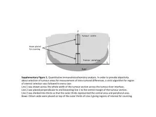
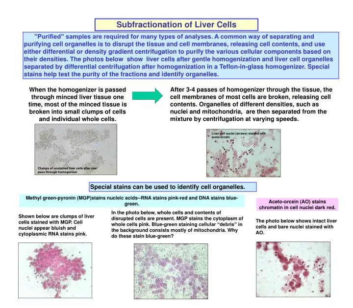
Liver cell nuclei (arrows) stained with aceto-orcein. Clumps of unstained liver cells after one pass through homogenizer. Subfractionation of Liver Cells.

E N D
Liver cell nuclei (arrows) stained with aceto-orcein Clumps of unstained liver cells after one pass through homogenizer Subfractionation of Liver Cells ”Purified” samples are required for many types of analyses. A common way of separating and purifying cell organelles is to disrupt the tissue and cell membranes, releasing cell contents, and use either differential or density gradient centrifugation to purify the various cellular components based on their densities. The photos below show liver cells after gentle homogenization and liver cell organelles separated by differential centrifugation after homogenization in a Teflon-in-glass homogenizer. Special stains help test the purity of the fractions and identify organelles. When the homogenizer is passed through minced liver tissue one time, most of the minced tissue is broken into small clumps of cells and individual whole cells. After 3-4 passes of homogenizer through the tissue, the cell membranes of most cells are broken, releasing cell contents. Organelles of different densities, such as nuclei and mitochondria, are then separated from the mixture by centrifugation at varying speeds. Special stains can be used to identify cell organelles. Methyl green-pyronin (MGP)stains nucleic acids--RNA stains pink-red and DNA stains blue-green. Aceto-orcein (AO) stains chromatin in cell nuclei dark red. In the photo below, whole cells and contents of disrupted cells are present. MGP stains the cytoplasm of whole cells pink. Blue-green staining cellular “debris” in the background consists mostly of mitochondria. Why do these stain blue-green? Shown below are clumps of liver cells stained with MGP. Cell nuclei appear bluish and cytoplasmic RNA stains pink. The photo below shows intact liver cells and bare nuclei stained with AO.