Download
1 / 29
511 likes | 1.06k Views
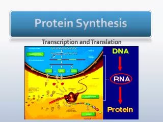
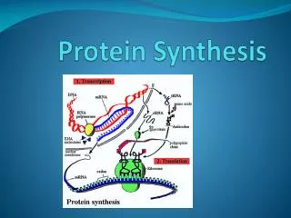
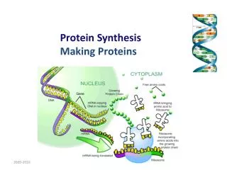
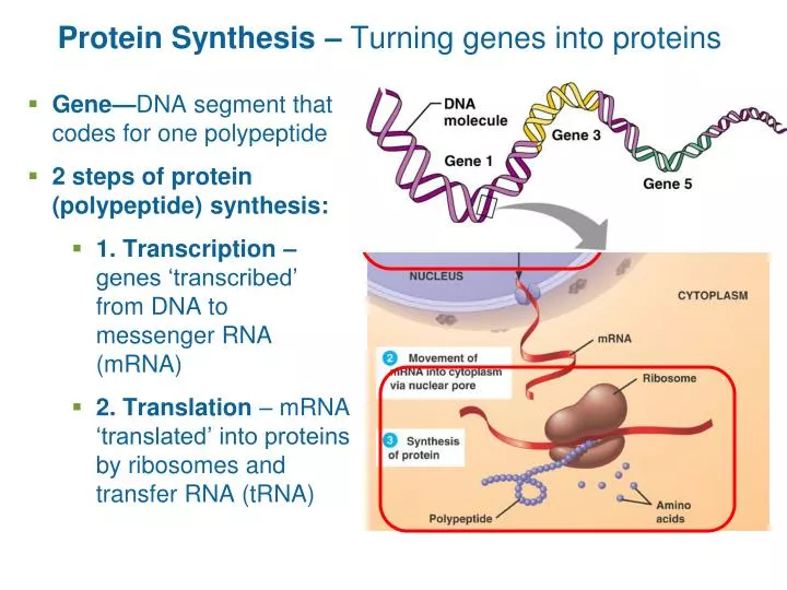
Protein Synthesis – Turning genes into proteins. Gene— DNA segment that codes for one polypeptide 2 steps of protein (polypeptide) synthesis: 1. Transcription – genes ‘transcribed’ from DNA to messenger RNA (mRNA)

E N D
Protein Synthesis – Turning genes into proteins • Gene—DNA segment that codes for one polypeptide • 2 steps of protein (polypeptide) synthesis: • 1. Transcription – genes ‘transcribed’ from DNA to messenger RNA (mRNA) • 2. Translation – mRNA ‘translated’ into proteins by ribosomes and transfer RNA (tRNA)
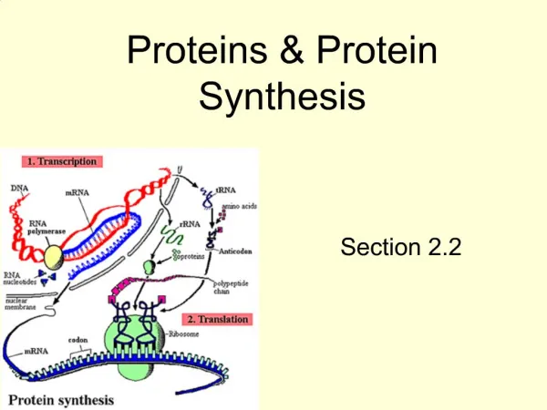
From DNA to Protein DNA Figure 3.33
From DNA to Protein DNA Transcription Figure 3.33
From DNA to Protein DNA Transcription Pre-mRNA RNA Processing mRNA Figure 3.33
From DNA to Protein Nuclear envelope DNA Transcription Pre-mRNA RNA Processing mRNA Figure 3.33
From DNA to Protein Nuclear envelope DNA Transcription Pre-mRNA RNA Processing mRNA Ribosome Translation Polypeptide Figure 3.33
Transcription: DNA genes mRNA • Transcription factors = proteins that… • Loosen histones from DNA • Bind to promoter = DNA sequence specifying start site of mRNA synthesis • Help bind RNA polymerase to promoter Figure 3.34
Nucleus Nuclear membrane RNA polymerase Nuclear pore mRNA Template strand of DNA Released mRNA RNA Polymerase = enzyme that makes mRNA • Unwinds DNA template • DNA bases (ATGC) pair with RNA bases (UACG) • A-U, T-A, C-G, G-C • Joins RNA nucleotides together • Releases at termination signal (specific set of DNA bases at end of gene) to stop transcription T A
Coding strand Termination signal Promoter Template strand Transcription unit In a process mediated by a transcription factor, RNA polymerase binds to promoter and unwinds 16–18 base pairs of the DNA template strand RNA polymerase Unwound DNA RNA polymerase bound to promoter RNA nucleotides mRNA synthesis begins mRNA RNA nucleotides RNA polymerase moves down DNA; mRNA elongates RNA polymerase mRNA synthesis is terminated DNA (a) mRNA transcript Coding strand RNA polymerase Unwinding of DNA Rewinding of DNA Template strand RNA nucleotides mRNA RNA-DNA hybrid region (b) Figure 3.34
The mRNA code is then translated into proteins • Codon – 3 bases of mRNA code for 1 amino acid • Processed mRNA leaves nucleus and binds with ribosome • Ribosome coordinates polypeptide construction with tRNA
Translation: mRNA proteins tRNA – brings amino acids to ribosomes Figure 3.16
enzyme tRNA ATP Three-dimensional structure Transfer RNA Molecules Serve as Interpreters During Translation • tRNA: single strand of RNA about 80 nucleotides in length • One single stranded loop = anticodon (special triplet of bases that pairs with complementary mRNA sequence) • At the other end, aa attachment site • Enzymes add amino acids to tRNA • (one enzyme per amino acid)
Nucleus Nuclear membrane RNA polymerase Nuclear pore mRNA Template strand of DNA Released mRNA Figure 3.36
Nucleus Nuclear membrane RNA polymerase Nuclear pore mRNA Template strand of DNA Released mRNA 1 After mRNA processing, mRNA leaves nucleus and attaches to ribosome, and translation begins. Small ribosomal subunit Codon 15 Codon 16 Codon 17 Direction of ribosome advance Portion of mRNA already translated Large ribosomal subunit Figure 3.36
Nucleus Nuclear membrane RNA polymerase Nuclear pore mRNA Template strand of DNA Amino acids Released mRNA 1 After mRNA processing, mRNA leaves nucleus and attaches to ribosome, and translation begins. tRNA Aminoacyl-tRNA synthetase Small ribosomal subunit Codon 15 Codon 16 Codon 17 Direction of ribosome advance Portion of mRNA already translated Large ribosomal subunit Energized by ATP, the correct amino acid is attached to each species of tRNA by aminoacyl-tRNA synthetase enzyme. Figure 3.36
Nucleus Nuclear membrane RNA polymerase Nuclear pore mRNA Template strand of DNA Amino acids Released mRNA 1 After mRNA processing, mRNA leaves nucleus and attaches to ribosome, and translation begins. tRNA Aminoacyl-tRNA synthetase Small ribosomal subunit Codon 15 Codon 16 Codon 17 Direction of ribosome advance Portion of mRNA already translated tRNA “head” bearing anticodon Large ribosomal subunit Energized by ATP, the correct amino acid is attached to each species of tRNA by aminoacyl-tRNA synthetase enzyme. 2 Incoming aminoacyl- tRNA hydrogen bonds via its anticodon to complementary mRNA sequence (codon) at the A site on the ribosome. Figure 3.36
Nucleus Nuclear membrane RNA polymerase Nuclear pore mRNA Template strand of DNA Amino acids Released mRNA 1 After mRNA processing, mRNA leaves nucleus and attaches to ribosome, and translation begins. tRNA Aminoacyl-tRNA synthetase Small ribosomal subunit Codon 15 Codon 16 Codon 17 Direction of ribosome advance Portion of mRNA already translated tRNA “head” bearing anticodon Large ribosomal subunit Energized by ATP, the correct amino acid is attached to each species of tRNA by aminoacyl-tRNA synthetase enzyme. 2 Incoming aminoacyl- tRNA hydrogen bonds via its anticodon to complementary mRNA sequence (codon) at the A site on the ribosome. 3 As the ribosome moves along the mRNA, a new amino acid is added to the growing protein chain and the tRNA in the A site is translocated to the P site. Figure 3.36
Nucleus Nuclear membrane RNA polymerase Nuclear pore mRNA Template strand of DNA Amino acids Released mRNA 1 After mRNA processing, mRNA leaves nucleus and attaches to ribosome, and translation begins. tRNA Aminoacyl-tRNA synthetase Small ribosomal subunit Codon 15 Codon 16 Codon 17 Direction of ribosome advance Portion of mRNA already translated tRNA “head” bearing anticodon Large ribosomal subunit Energized by ATP, the correct amino acid is attached to each species of tRNA by aminoacyl-tRNA synthetase enzyme. 2 Incoming aminoacyl- tRNA hydrogen bonds via its anticodon to complementary mRNA sequence (codon) at the A site on the ribosome. 3 As the ribosome moves along the mRNA, a new amino acid is added to the growing protein chain and the tRNA in the A site is translocated to the P site. 4 Once its amino acid is released, tRNA is ratcheted to the E site and then released to reenter the cytoplasmic pool, ready to be recharged with a new amino acid. Figure 3.36
At the ribosome… P site on ribosome holds tRNA that holds growing polypeptide A site on ribosome receives next tRNA w/ next amino acid Amino acid is added to polypeptide tRNA shift positions from A to P after amino acids are added
Amino Acids Added Until Stop Codon Terminates Translation Ribosomes disassemble and start translating another mRNA
Information Transfer from DNA to RNA to polypeptide Figure 3.38
Cells differentiate by turning on different genes (i.e. making different proteins) Different genes turned ‘on’ or ‘off’ by chemical signals Early development likely controlled by simple gradients of CO2 and O2
Cytosol mRNA 1 Signal sequence Receptor site Signal- recognition particle (SRP) ER cisterna ER membrane Signal Mechanism of Protein Synthesis Ribosomes of rough ER are transient (i.e. not permanently attached) Signal sequence = short amino acid chain of new protein that binds to an SRP (signal recognition particle) Figure 3.19
Cytosol mRNA 2 1 Signal sequence Receptor site Growing polypeptide Signal- recognition particle (SRP) ER cisterna ER membrane Signal Mechanism of Protein Synthesis SRP guides ribosome – polypeptide (protein) complex to ER by binding to receptors on rough ER SRP removed by enzymes at receptor site Figure 3.19
Cytosol Ribosomes mRNA 3 2 1 Signal sequence removed Signal sequence Receptor site Growing polypeptide Signal- recognition particle (SRP) ER cisterna ER membrane Signal Mechanism of Protein Synthesis Original signal sequence quickly removed as well Protein continues forming inside ER (sometimes sugars added) Figure 3.19
Cytosol Ribosomes mRNA 3 4 2 1 Released glycoprotein Signal sequence removed Signal sequence Receptor site Growing polypeptide Signal- recognition particle (SRP) ER cisterna ER membrane Signal Mechanism of Protein Synthesis Primary structure completes, ribosomes detach Folding of proteins often aided by chaperones Integral proteins remain embedded in membrane Figure 3.19
Cytosol Transport vesicle budding off 5 Ribosomes mRNA 3 4 Sugar group 2 1 Released glycoprotein Signal sequence removed Signal sequence Receptor site Growing polypeptide Signal- recognition particle (SRP) ER cisterna ER membrane Signal Mechanism of Protein Synthesis Coatomer-coated transport vesicle breaks away Coatomer = proteins that aid intracellular vesicle formation Figure 3.19
Cytosol Transport vesicle budding off Coatomer- coated transport vesicle 5 Ribosomes mRNA 3 4 Sugar group 2 1 Released glycoprotein Signal sequence removed Signal sequence Receptor site Growing polypeptide Signal- recognition particle (SRP) ER cisterna ER membrane Signal Mechanism of Protein Synthesis Where does this go? Figure 3.19
Rough ER Cisterna Proteins in cisterna Phagosome Membrane Vesicle Lysosomes containing acid hydrolase enzymes Vesicle incorporated into plasma membrane Pathway 3 Coatomer coat Golgi apparatus Pathway 2 Secretory vesicles Pathway 1 Plasma membrane Proteins Secretion by exocytosis Extracellular fluid Golgi receives from its cis side, ships from the trans side Figure 3.21