Download
1 / 51
510 likes | 605 Views
The Human Immune System is an excellent example of variety in cell structure and function. Basic Information. In order to understand how these cells function, we need to have a general understanding of the immune system itself. Basic Information.

E N D
The Human Immune System is an excellent example of variety in cell structure and function
BasicInformation In order to understand how these cells function, we need to have a general understanding of the immune system itself.
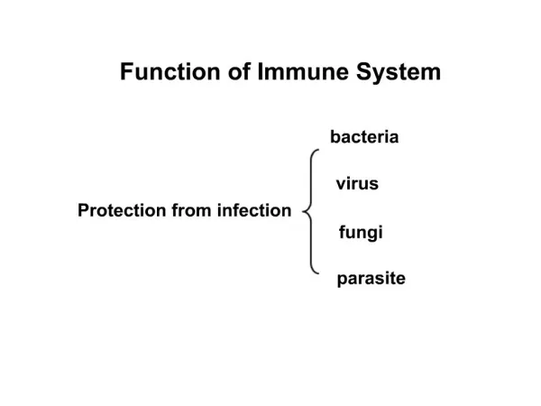
BasicInformation In order to understand how these cells function, we need to have a general understanding of the immune system itself. The job of your immune system is to keep foreign substances, usually called pathogens, from invading and infecting your body.
BasicInformation In order to understand how these cells function, we need to have a general understanding of the immune system itself. The job of your immune system is to keep foreign substances, usually called pathogens, from invading and infecting your body. One of the major challenges of the immune system is to be able to quickly and efficiently identify your own (“self”) cells so that only pathogens are targeted.
BasicInformation In order to understand how these cells function, we need to have a general understanding of the immune system itself. The job of your immune system is to keep foreign substances, usually called pathogens, from invading and infecting your body. One of the major challenges of the immune system is to be able to quickly and efficiently identify your own (“self”) cells so that only pathogens are targeted. A number of different organs and tissues are needed for this to occur.
The Phagocytes Phagocytes are the soldiers of the immune system, and provide innate immunity. They are responsible for swallowing, killing and digesting invading microbes. The process of swallowing microbes is known as phagocytosis. There are two main types of phagocyte * Microphages. These cells are also known as Polymorphonuclear Leucocytes, PMNs and Polymorphs. These cells start life in the bone marrow. They are constantly circulating in the blood. They cannot replicate, and live for only a few days. The bone marrow contains large reserves of microphages. * Macrophages. These cells start out life as monocytes, which originate in the stem cells in the bone marrow, but when they are first called into action, they turn into macrophages. Macrophages are not as numerous as microphages, and there are no large reserves of them, but they are longer lived than microphages. Macrophages are stationed at strategic locations throughout the body, usually in places that are not otherwise well defended. These areas include the alveoli of the lungs, the abdominal (peritoneal) and chest (pleural) cavities, under the top layer of the skin and the intestines. Macrophages are the front line of defense against microbial invasion in these areas. As mentioned above, the process of swallowing of microbes by the phagocytes is known as phagocytosis. After the invading microbe has been swallowed, the next task for the phagocyte is to kill the microbe. This is achieved in two main ways. * Aerobically, i.e. using oxygen. The phagocytes produce oxygen based chemicals that are highly disruptive to the swallowed microbe. Oxygen is highly chemically reactive, and these oxygen based chemicals "tear" the microbe apart. This process is known as the oxidative burst, or the respiratory burst. * Anaerobically, i.e. without using oxygen. One way to kill the microbe without oxygen is by using a chemical that deprives the microbe of iron, thus preventing it from metabolising. Another way is to increase the acidity of the internal environment of the phagocyte. When these tasks are complete, the Macrophages have one further task to complete. They return to the lymph nodes, displaying the remnants of the destroyed invader on their surface. This has the effect of stimulating the cells of the Acquired immunity system into action. If a pathogen can get past these barriers, the body must identify and remove it.
The Phagocytes Phagocytes are the soldiers of the immune system, and provide innate immunity. They are responsible for swallowing, killing and digesting invading microbes. The process of swallowing microbes is known as phagocytosis. There are two main types of phagocyte * Microphages. These cells are also known as Polymorphonuclear Leucocytes, PMNs and Polymorphs. These cells start life in the bone marrow. They are constantly circulating in the blood. They cannot replicate, and live for only a few days. The bone marrow contains large reserves of microphages. * Macrophages. These cells start out life as monocytes, which originate in the stem cells in the bone marrow, but when they are first called into action, they turn into macrophages. Macrophages are not as numerous as microphages, and there are no large reserves of them, but they are longer lived than microphages. Macrophages are stationed at strategic locations throughout the body, usually in places that are not otherwise well defended. These areas include the alveoli of the lungs, the abdominal (peritoneal) and chest (pleural) cavities, under the top layer of the skin and the intestines. Macrophages are the front line of defense against microbial invasion in these areas. As mentioned above, the process of swallowing of microbes by the phagocytes is known as phagocytosis. After the invading microbe has been swallowed, the next task for the phagocyte is to kill the microbe. This is achieved in two main ways. * Aerobically, i.e. using oxygen. The phagocytes produce oxygen based chemicals that are highly disruptive to the swallowed microbe. Oxygen is highly chemically reactive, and these oxygen based chemicals "tear" the microbe apart. This process is known as the oxidative burst, or the respiratory burst. * Anaerobically, i.e. without using oxygen. One way to kill the microbe without oxygen is by using a chemical that deprives the microbe of iron, thus preventing it from metabolising. Another way is to increase the acidity of the internal environment of the phagocyte. When these tasks are complete, the Macrophages have one further task to complete. They return to the lymph nodes, displaying the remnants of the destroyed invader on their surface. This has the effect of stimulating the cells of the Acquired immunity system into action. If a pathogen can get past these barriers, the body must identify and remove it. -- The skin and the lining of the body cavities that open to the outside must provide a protective barrier.
The Phagocytes Phagocytes are the soldiers of the immune system, and provide innate immunity. They are responsible for swallowing, killing and digesting invading microbes. The process of swallowing microbes is known as phagocytosis. There are two main types of phagocyte * Microphages. These cells are also known as Polymorphonuclear Leucocytes, PMNs and Polymorphs. These cells start life in the bone marrow. They are constantly circulating in the blood. They cannot replicate, and live for only a few days. The bone marrow contains large reserves of microphages. * Macrophages. These cells start out life as monocytes, which originate in the stem cells in the bone marrow, but when they are first called into action, they turn into macrophages. Macrophages are not as numerous as microphages, and there are no large reserves of them, but they are longer lived than microphages. Macrophages are stationed at strategic locations throughout the body, usually in places that are not otherwise well defended. These areas include the alveoli of the lungs, the abdominal (peritoneal) and chest (pleural) cavities, under the top layer of the skin and the intestines. Macrophages are the front line of defense against microbial invasion in these areas. As mentioned above, the process of swallowing of microbes by the phagocytes is known as phagocytosis. After the invading microbe has been swallowed, the next task for the phagocyte is to kill the microbe. This is achieved in two main ways. * Aerobically, i.e. using oxygen. The phagocytes produce oxygen based chemicals that are highly disruptive to the swallowed microbe. Oxygen is highly chemically reactive, and these oxygen based chemicals "tear" the microbe apart. This process is known as the oxidative burst, or the respiratory burst. * Anaerobically, i.e. without using oxygen. One way to kill the microbe without oxygen is by using a chemical that deprives the microbe of iron, thus preventing it from metabolising. Another way is to increase the acidity of the internal environment of the phagocyte. When these tasks are complete, the Macrophages have one further task to complete. They return to the lymph nodes, displaying the remnants of the destroyed invader on their surface. This has the effect of stimulating the cells of the Acquired immunity system into action. If a pathogen can get past these barriers, the body must identify and remove it. -- The skin and the lining of the body cavities that open to the outside must provide a protective barrier. --The entrance to the organs like the gut and the reproductive tract needs to prevent invasion by any pathogenic micro organisms.
The Phagocytes Phagocytes are the soldiers of the immune system, and provide innate immunity. They are responsible for swallowing, killing and digesting invading microbes. The process of swallowing microbes is known as phagocytosis. There are two main types of phagocyte * Microphages. These cells are also known as Polymorphonuclear Leucocytes, PMNs and Polymorphs. These cells start life in the bone marrow. They are constantly circulating in the blood. They cannot replicate, and live for only a few days. The bone marrow contains large reserves of microphages. * Macrophages. These cells start out life as monocytes, which originate in the stem cells in the bone marrow, but when they are first called into action, they turn into macrophages. Macrophages are not as numerous as microphages, and there are no large reserves of them, but they are longer lived than microphages. Macrophages are stationed at strategic locations throughout the body, usually in places that are not otherwise well defended. These areas include the alveoli of the lungs, the abdominal (peritoneal) and chest (pleural) cavities, under the top layer of the skin and the intestines. Macrophages are the front line of defense against microbial invasion in these areas. As mentioned above, the process of swallowing of microbes by the phagocytes is known as phagocytosis. After the invading microbe has been swallowed, the next task for the phagocyte is to kill the microbe. This is achieved in two main ways. * Aerobically, i.e. using oxygen. The phagocytes produce oxygen based chemicals that are highly disruptive to the swallowed microbe. Oxygen is highly chemically reactive, and these oxygen based chemicals "tear" the microbe apart. This process is known as the oxidative burst, or the respiratory burst. * Anaerobically, i.e. without using oxygen. One way to kill the microbe without oxygen is by using a chemical that deprives the microbe of iron, thus preventing it from metabolising. Another way is to increase the acidity of the internal environment of the phagocyte. When these tasks are complete, the Macrophages have one further task to complete. They return to the lymph nodes, displaying the remnants of the destroyed invader on their surface. This has the effect of stimulating the cells of the Acquired immunity system into action. If a pathogen can get past these barriers, the body must identify and remove it. -- The skin and the lining of the body cavities that open to the outside must provide a protective barrier. --The entrance to the organs like the gut and the reproductive tract needs to prevent invasion by any pathogenic micro organisms. --The mucosal membranes secrete a variety of fluids, such as saliva by the intestinal tract and mucus in the respiratory tract, which provide a defense against foreign micro-organisms.
The Phagocytes Phagocytes are the soldiers of the immune system, and provide innate immunity. They are responsible for swallowing, killing and digesting invading microbes. The process of swallowing microbes is known as phagocytosis. There are two main types of phagocyte * Microphages. These cells are also known as Polymorphonuclear Leucocytes, PMNs and Polymorphs. These cells start life in the bone marrow. They are constantly circulating in the blood. They cannot replicate, and live for only a few days. The bone marrow contains large reserves of microphages. * Macrophages. These cells start out life as monocytes, which originate in the stem cells in the bone marrow, but when they are first called into action, they turn into macrophages. Macrophages are not as numerous as microphages, and there are no large reserves of them, but they are longer lived than microphages. Macrophages are stationed at strategic locations throughout the body, usually in places that are not otherwise well defended. These areas include the alveoli of the lungs, the abdominal (peritoneal) and chest (pleural) cavities, under the top layer of the skin and the intestines. Macrophages are the front line of defense against microbial invasion in these areas. As mentioned above, the process of swallowing of microbes by the phagocytes is known as phagocytosis. After the invading microbe has been swallowed, the next task for the phagocyte is to kill the microbe. This is achieved in two main ways. * Aerobically, i.e. using oxygen. The phagocytes produce oxygen based chemicals that are highly disruptive to the swallowed microbe. Oxygen is highly chemically reactive, and these oxygen based chemicals "tear" the microbe apart. This process is known as the oxidative burst, or the respiratory burst. * Anaerobically, i.e. without using oxygen. One way to kill the microbe without oxygen is by using a chemical that deprives the microbe of iron, thus preventing it from metabolising. Another way is to increase the acidity of the internal environment of the phagocyte. When these tasks are complete, the Macrophages have one further task to complete. They return to the lymph nodes, displaying the remnants of the destroyed invader on their surface. This has the effect of stimulating the cells of the Acquired immunity system into action. If a pathogen can get past these barriers, the body must identify and remove it. -- The skin and the lining of the body cavities that open to the outside must provide a protective barrier. --The entrance to the organs like the gut and the reproductive tract needs to prevent invasion by any pathogenic micro organisms. --The mucosal membranes secrete a variety of fluids, such as saliva by the intestinal tract and mucus in the respiratory tract, which provide a defense against foreign micro-organisms. -- The body carries its own natural microorganisms that we happily live with, which also prevent other more dangerous bugs from taking over. --Adapted from www.julies-story.org
Blood There are three major components to human blood.
Blood There are three major components to human blood. Human blood is approximately 55% plasma, which is the “fluid” part of the blood with ions, proteins and other substances dissolved in it.
Blood There are three major components to human blood. Human blood is approximately 55% plasma, which is the “fluid” part of the blood with ions, proteins and other substances dissolved in it. Image:http://www.nursing.ucla.edu/Userpages/mwoo/cbc/smear.htm
Blood There are three major components to human blood. Human blood is approximately 55% plasma, which is the “fluid” part of the blood with ions, proteins and other substances dissolved in it. Image:http://www.nursing.ucla.edu/Userpages/mwoo/cbc/smear.htm
Blood There are three major components to human blood. Human blood is approximately 55% plasma, which is the “fluid” part of the blood with ions, proteins and other substances dissolved in it. The “cellular elements” blood make up the other 45%. Almost 95% of these are red blood cells (erythrocytes) that carry oxygen in the blood. Image:http://www.nursing.ucla.edu/Userpages/mwoo/cbc/smear.htm
Blood There are three major components to human blood. Human blood is approximately 55% plasma, which is the “fluid” part of the blood with ions, proteins and other substances dissolved in it. The “cellular elements” blood make up the other 45%. Almost 95% of these are red blood cells (erythrocytes) that carry oxygen in the blood. Image:http://www.nursing.ucla.edu/Userpages/mwoo/cbc/smear.htm
Blood There are three major components to human blood. Human blood is approximately 55% plasma, which is the “fluid” part of the blood with ions, proteins and other substances dissolved in it. The “cellular elements” blood make up the other 45%. Almost 95% of these are red blood cells (erythrocytes) that carry oxygen in the blood. About 5% of the cellular elements in blood are platelets. These are cell pieces that are used for blood clotting. Image:http://www.nursing.ucla.edu/Userpages/mwoo/cbc/smear.htm
Blood There are three major components to human blood. Human blood is approximately 55% plasma, which is the “fluid” part of the blood with ions, proteins and other substances dissolved in it. The “cellular elements” blood make up the other 45%. Almost 95% of these are red blood cells (erythrocytes) that carry oxygen in the blood. About 5% of the cellular elements in blood are platelets. These are cell pieces that are used for blood clotting. Image:http://www.nursing.ucla.edu/Userpages/mwoo/cbc/smear.htm
Blood There are three major components to human blood. Human blood is approximately 55% plasma, which is the “fluid” part of the blood with ions, proteins and other substances dissolved in it. The “cellular elements” blood make up the other 45%. Almost 95% of these are red blood cells (erythrocytes) that carry oxygen in the blood. About 5% of the cellular elements are platelets. These are cell pieces that are used for blood clotting. Much less than 1% of blood contains white blood cells, (leukocytes), they are vitally important in fighting infection. Image:http://www.nursing.ucla.edu/Userpages/mwoo/cbc/smear.htm
Blood There are three major components to human blood. Human blood is approximately 55% plasma, which is the “fluid” part of the blood with ions, proteins and other substances dissolved in it. The “cellular elements” blood make up the other 45%. Almost 95% of these are red blood cells (erythrocytes) that carry oxygen in the blood. About 5% of the cellular elements are platelets. These are cell pieces that are used for blood clotting. Much less than 1% of blood contains white blood cells, (leukocytes), they are vitally important in fighting infection. Image:http://www.nursing.ucla.edu/Userpages/mwoo/cbc/smear.htm
Cell Sizes The red blood cells are approximately 8 um across and are generally very regular in their size and shape. Image:http://www.nursing.ucla.edu/Userpages/mwoo/cbc/smear.htm
Cell Sizes The red blood cells are approximately 8 um across and are generally very regular in their size and shape. Platelets are about one third to one half as large as red blood cells, about 2-4 um across. Image:http://www.nursing.ucla.edu/Userpages/mwoo/cbc/smear.htm
Cell Sizes The red blood cells are approximately 8 um across and are generally very regular in their size and shape. Platelets are about one third to one half as large as red blood cells, about 2-4 um across. White blood cells are often larger than the red cells, generally 9 - 12 um across. This measurement may vary a great deal since there are many different types of white blood cells. Image:http://www.nursing.ucla.edu/Userpages/mwoo/cbc/smear.htm
Cell Sizes For comparison, let’s look at a photograph of a human cheek cell (~50 um) shown at the same scale as our blood cells:
Cell Sizes For comparison, let’s look at a photograph of a human cheek cell (~50 um) shown at the same scale as our blood cells: Image:http://www.nursing.ucla.edu/Userpages/mwoo/cbc/smear.htm Image:http://www.cat.cc.md.us/courses/bio141/lecguide/unit1/prostruct/euproreview/epit.html
Cell Sizes For comparison, let’s look at a photograph of a human cheek cell (~50 um) shown at the same scale as our blood cells: Image:http://www.nursing.ucla.edu/Userpages/mwoo/cbc/smear.htm Here you can see the stained nucleus and the small, darkly stained bacteria that are all over the surface of the cheek cell. Image:http://www.cat.cc.md.us/courses/bio141/lecguide/unit1/prostruct/euproreview/epit.html
Cell Sizes For comparison, let’s look at a photograph of a human cheek cell (~50 um) shown at the same scale as our blood cells: Image:http://www.nursing.ucla.edu/Userpages/mwoo/cbc/smear.htm Here you can see the stained nucleus and the small, darkly stained bacteria that are all over the surface of the cheek cell. Notice that typical blood cells are smaller than even the nucleus of a cheek cell. Image:http://www.cat.cc.md.us/courses/bio141/lecguide/unit1/prostruct/euproreview/epit.html
Cell Sizes For comparison, let’s look at a photograph of a human cheek cell (~50 um) shown at the same scale as our blood cells: Image:http://www.nursing.ucla.edu/Userpages/mwoo/cbc/smear.htm Here you can see the stained nucleus and the small, darkly stained bacteria that are all over the surface of the cheek cell. Notice that typical blood cells are smaller than even the nucleus of a cheek cell. Image:http://www.cat.cc.md.us/courses/bio141/lecguide/unit1/prostruct/euproreview/epit.html
There are 5 major types of white blood cells (leukocytes). All of them play an important role in fighting disease.
There are 5 major types of white blood cells (leukocytes). All of them play an important role in fighting disease. 1. Neutrophils leave the blood to go to tissues where infection or inflammation is developing. They mainly engulf and destroy bacteria and fungi.
There are 5 major types of white blood cells (leukocytes). All of them play an important role in fighting disease. 1. Neutrophils leave the blood to go to tissues where infection or inflammation is developing. They mainly engulf and destroy bacteria and fungi. Normal Red Blood Cell
There are 5 major types of white blood cells (leukocytes). All of them play an important role in fighting disease. 1. Neutrophils leave the blood to go to tissues where infection or inflammation is developing. They mainly engulf and destroy bacteria and fungi. Normal Red Blood Cell Neutrophil
There are 5 major types of white blood cells (leukocytes). 2. Eosinophils attack organisms that are too big to be eaten by a single phagocyte, like worms.
There are 5 major types of white blood cells (leukocytes). 2. Eosinophils attack organisms that are too big to be eaten by a single phagocyte, like worms. This image shows red blood cells [R], a neutrophil [N] and an eosinophil [E]. Image:http://www.cytochemistry.net/microanatomy/blood/blood_cells.htm#RED%20BLOOD%20CELLS
There are 5 major types of white blood cells (leukocytes). 3. Basophils do not attack and “swallow” invading cells; they release chemical that help the body’s allergic response to a pathogen.
There are 5 major types of white blood cells (leukocytes). 3. Basophils do not attack and “swallow” invading cells; they release chemical that help the body’s allergic response to a pathogen. Basophil surrounded by red blood cells.
There are 5 major types of white blood cells (leukocytes). 4. Monocytes are cells released into the blood from the bone marrow. When they get to a particular site in an organism they may change into macrophages that engulf and destroy invading pathogens.
There are 5 major types of white blood cells (leukocytes). 4. Monocytes are cells released into the blood from the bone marrow. When they get to a particular site in an organism they may change into macrophages that engulf and destroy invading pathogens. Red blood cells Monocyte Image: http://image.bloodline.net/stories/storyReader$1628
There are 5 major types of white blood cells (leukocytes). 5. Lymphocytes are the fifth group of white blood cells; they are divided into three categories:
There are 5 major types of white blood cells (leukocytes). 5. Lymphocytes are the fifth group of white blood cells; they are divided into three categories: -Natural killer cells attack tumor cells and some cells that have been infected with viruses.
There are 5 major types of white blood cells (leukocytes). 5. Lymphocytes are the fifth group of white blood cells; they are divided into three categories: -Natural killer cells attack tumor cells and some cells that have been infected with viruses. -B-lymphocytes develop in the bone marrow.
There are 5 major types of white blood cells (leukocytes). 5. Lymphocytes are the fifth group of white blood cells; they are divided into three categories: -Natural killer cells attack tumor cells and some cells that have been infected with viruses. -B-lymphocytes develop in the bone marrow. -T-lymphocytes develop in the thymus.
There are 5 major types of white blood cells (leukocytes). 5. Lymphocytes are the fifth group of white blood cells; they are divided into three categories: -Natural killer cells attack tumor cells and some cells that have been infected with viruses. -B-lymphocytes develop in the bone marrow. -T-lymphocytes develop in the thymus. Lymphocytes originate in the bone marrow, but can proliferate in the spleen, thymus and other lymphoid tissues. Often, large lymphocytes seen in the blood have been activated somewhere in the body, and are traveling to sites of action. Image:http://oac.med.jhmi.edu/pathconcepts/ShowImage.cfm?TutorialID=7&ConceptID=27&ImageID=259
There are 5 major types of white blood cells (leukocytes). 5. Lymphocytes are the fifth group of white blood cells; they are divided into three categories: -Natural killer cells attack tumor cells and some cells that have been infected with viruses. -B-lymphocytes develop in the bone marrow. -T-lymphocytes develop in the thymus. Both B- and T- cells are covered with many different molecules. If one of these matches up with a molecule of a pathogen, the B- or T-cell may engulf the pathogen and destroy it. Then the body can make many, many copies of this cell to fight the pathogen.
There are 5 major types of white blood cells (leukocytes). 5. Lymphocytes are the fifth group of white blood cells; they are divided into three categories: -Natural killer cells attack tumor cells and some cells that have been infected with viruses. -B-lymphocytes develop in the bone marrow. -T-lymphocytes develop in the thymus. Both B- and T- cells are covered with many different molecules. If one of these matches up with a molecule of a pathogen, the B- or T-cell may engulf the pathogen and destroy it. Then the body can make many, many copies of this cell to fight the pathogen. The body keeps a “memory” of every B- or T- cell that has been activated and it is able to attack that particular foreign body almost instantly if it appears again.
Why should I care about the immune system? Like many parts of the body, we learn a great deal about the immune system by studying what happens when it doesn’t work properly.
Why should I care about the immune system? Like many parts of the body, we learn a great deal about the immune system by studying what happens when it doesn’t work properly. Sometimes the body is no longer able to recognize certain normally occurring cell types. When this happens the immune system identifies these as foreign cells and begins to attack them. This results in an autoimmune disease.
Why should I care about the immune system cells? Like many parts of the body, we learn a great deal about the immune system by studying what happens when it doesn’t work properly. Sometimes the body is no longer able to recognize certain normally occurring cell types. When this happens the immune system identifies these as foreign cells and begins to attack them. This results in an autoimmune disease. Of the scores of autoimmune diseases the have been discovered, some of the more common are: