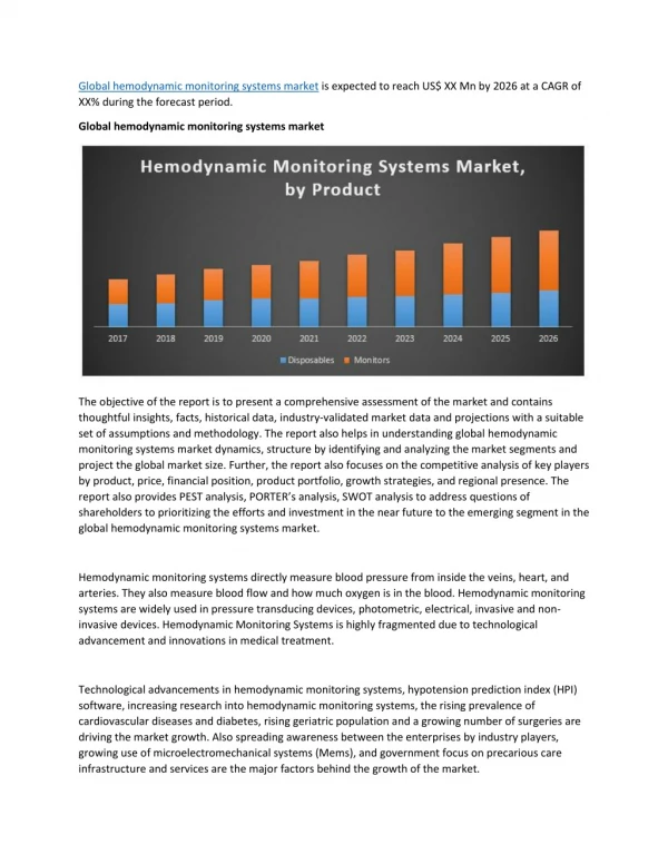
Download

1 / 36
370 likes | 435 Views
Physiologic Basis for Hemodynamic Monitoring. 臺大醫院麻醉部 鄭雅蓉. Circulation to Perfusion. Oxygenation Consumption. Arteries. Sympathetic Nervous System. Heart. Organs & Tissues. Veins. Anesthesia Sedation. Adequate Oxygen Delivery?. Demand. Consumption. Oxygen Delivery.

E N D
Physiologic Basis for Hemodynamic Monitoring • 臺大醫院麻醉部 • 鄭雅蓉
Circulation to Perfusion Oxygenation Consumption Arteries Sympathetic Nervous System Heart Organs & Tissues Veins Anesthesia Sedation
Adequate Oxygen Delivery? Demand Consumption
Oxygen Delivery Hemodynamic Monitors Oxygen Delivery Cardiac Output Oxygen Content = X Arterial Blood Gas Hemoglobin PaO2 Oxygen Content
Oxygen Consumption Oxygen Delivery Oxygen Consumed Remaining Oxygen to Heart = + Oxygen Uptake by Organs & Tissues Oxygen Content in CVP & PA
Physiological Truth • There is no such thing as a “Normal Cardiac Output” • Cardiac output is either • Absolute values can only be used as minimal levels below which some tissue beds are probably under perfused - Adequate to meet the metabolic demands - Inadequate to meet the metabolic demands
History of Monitoring Pressure, arterial line & CVP • 1960s: golden age of vasopressors • 1970s: golden age of inotropes • 1980s: • 1990s till now: Cardiac output, PA catheter SvO2 , relative balance between oxygen supply and demand Better understanding of tissue oxygenation, right ventricular function Functional monitoring, PiCCO, continuous CO Less invasive, TEE
Hemodynamic Monitoring Truth • No monitoring device, no matter how simple or complex, invasive or non-invasive, inaccurate or precise will improve outcome • Unless coupled to a treatment, which itself improves outcome Pinsky & Payen. Functional Hemodynamic Monitoring, Springer, 2004
Goals of Monitors • To assure the adequacy of perfusion • Early detection of inadequacy of perfusion • To titrate therapy to specific hemodynamic end point • To differentiate among various organ system dysfunctions Hemodynamic monitoring for individual patient should be physiologically based and goal oriented.
Different Environments Demand Different Rules • Emergency Department • Trauma ICU • Operation Room • ICU & RR Rapid, minimally invasive, high sensitivity Rapid, invasive, high specificity Accurate, invasive, high specificity Close titration, zero tolerance for complications Somewhere in between ER and OR
Hemodynamic monitors (1) • Traditional invasive monitors • Arterial line • CVP & ScvO2 • PA catheter, CCO, SvO2 • Functional pressure variation • Pulse pressure variation • Stroke volume variation
Hemodynamic monitors (2) • Alternative to right-side heart catheterization • PiCCO • Echocardiography • Transesophageal echocardiography (TEE) • Esophageal doppler monitor
Is Cardiac Output Adequate? Is blood flow adequate to meet metabolic demands? Pump function ? Adequate intravascular volume? Driving pressure for venous return?
Is Cardiac Output Adequate? We Should Know The effects of respiration or mechanical ventilation Left & right ventricular function Preload & preload responsiveness
Ventricular Function • Left ventricular function • Right ventricular function • Depressed right ventricular function was further linked to more severely compromised left ventricular function. Nielsen et al. Intensive care med 32:585-94, 2006
Respiration and RV function • Spontaneous ventilation • Mechanical positive pressure ventilation
Use of Heart Lung Interactions to Diagnose Preload-Responsiveness • ValSalva maneuver • Ventilation-induced changes in: • Right atrial pressure • Systolic arterial pressure • Arterial pulse pressure • Inferior vena caval diameter • Superior vena caval diameter Sharpey-Schaffer. Br Med J 1:693-699, 1955 Zema et al., D Chest 85,59-64, 1984 Magder et al. J Crit Care 7:76‑85, 1992 Perel et al. Anesthesiology 67:498-502, 1987 Michard et al. Am J Respir Crit Care Med 162:134-8, 2000 Jardin & Vieillard-Baron. Intensive Care Med 29:1426-34, 2003 Vieillard-Baron et al. Am J Respir Crit Care Med 168: 671-6, 2003
Mechanical positive pressure ventilation • Increase RV outflow impedance, reduce ejection, increase RVEDV, tricuspid regurgitation • TEE: SVC diameter: the effect of venous return? • CVP may be misleading
Preload & Preload Responsiveness • Starling’s law is still operated. • CVP, PAOP and their changes: If end diastolic volume ( EDV ) increased in response to volume loading, then stroke volume increased as well. Did not respond with EDV, but Provide a stable route for drug titration and fluid infusion
Neither CVP or Ppao reflect Ventricular Volumes or tract preload-responsiveness Kumar et al. Crit Care Med 32:691-9, 2004
Neither CVP or Ppao reflect Ventricular Volumes or tract preload-responsiveness Kumar et al. Crit Care Med 32:691-9, 2004
Physiological limitations CVP RV dysfunction Pulmonary hypertension LV dysfunction Tamponade & hyperinflation Intravascular volume expansion PAOP LV diastolic compliance Pericardial restraint Intrathoracic pressure Heart rate Mitral valvulopathy
Predicting Fluid Responsiveness in ICU Patients Michard & Teboul. Chest 121:2000-8, 2002
Can CVP Be Use for Fluid Management? • Relatively • Absolutely • Does apneic CVP predict preload responsiveness? Yes on most counts Yes for hypovolemia (10 mmHg cut-off) No, but then neither does Ppao or direct measures of LV end-diastolic volume Michard et al. Am J Respir Crit Care Med 162:134-8, 2000
Thermodilution Cardiac Output • Mean (steady state) blood flow • Functional significance of a specific cardiac output value • Cardiac output varies to match the metabolic demands of the body The meaning of cardiac output Pinsky, The meaning of cardiac output. Intensive Care Med 16:415-417, 1990
Mixed Venous Oximetry • SvO2 is the averaged end-capillary oxygen content (essential for VO2 Fick) • SvO2 is a useful parameter of hemodynamic status is specific conditions • If SvO2 < 60% some capillary beds ischemic • In sedated, paralyzed patient SvO2 parallels CO
Adequate Oxygen delivery? • SvO2: mixed venous oxygen saturation • C(a-v)O2: arterial-venous oxygen content difference • Lactate: the demand and need of the use of oxygen Consumption & delivery Consumption & cardiac output Consumption & demand
Central Venous and Mixed Venous O2 Saturation • ScvO2 on CVP monitor • SvO2 on PA catheter • SvO2 is a sensitive but non-specific measure of cardiovascular instability • Although ScvO2 tracked SvO2, it is tended to 7 ± 4 % higher.
Arterial Catheterization • Directly measured arterial blood pressure • Baroreceptor mechanisms defend arterial pressure over a wide range of flows • Hypotension is always pathological • Beat-to-beat variations in pulse pressure reflect changes in stroke volume rather than cardiac output
Pulmonary Arterial Catheterization • Pressures reflect intrathoracic pressure • Ventilation alters both pulmonary blood flow and vascular resistance • Resistance increases with increasing lung volume above resting lung volume (FRC) • Right ventricular output varies in phase with respiration-induced changes in venous return • Spontaneous inspiration increases pulmonary blood flow • Positive-pressure inspiration decreases pulmonary blood flow
Functional Hemodynamic Monitors • Arterial pulse contour analysis • A better monitors for preload responsiveness: • a significant correlation between the increase of cardiac index by fluid loading by pulse pressure variation and stroke volume variation • Peripheral continuous cardiac output system (PiCCO): arterial pulse contour and transpulmonary thermal injection: • intrathoracic volume and extravascular lung water
Conclusions Regarding Different Monitors • Hemodynamic monitoring becomes more effective at predicting cardiovascular function when measured using performance parameters • CVP and arterial pulse pressure (ΔPP) variations predict preload responsiveness • CVP, ScvO2 and PAOP, SvO2 predict the adequacy of oxygen transport
The Truths in Hemodynamics • Tachycardia is never a good thing. • Hypotension is always pathological. • There is no normal cardiac output. • CVP is only elevated in disease. • A higher mortality was shown in patients with right ventricular dysfunction and an increase of pulmonary vascular resistance.
The Truths in Hemodynamic Monitoring • Monitors associate with inaccuracies, misconceptions and poorly documented benefits. • A good understanding of the pathophysiological underpinnings for its effective application across patient groups is required. • Functional hemodynamic monitors are superior to conventional filling pressure. • The goal of treatments based on monitoring is to restore the physiological homeostasis.