Download

1 / 40
571 likes | 1.74k Views
STRABISMUS. DONE BY: Kamal Sub- Laban Mahmoud Salman Mustafa Jaber. Anatomy Review. The eye layers: 1- A tough outer coat (fibrous) . 2- A rich vascular coat (the choroid ) 3- innermost neural (retnia). Extra-ocular muscles. Medial and lateral recti horizontal eye movement

E N D
STRABISMUS DONE BY: Kamal Sub-Laban MahmoudSalman Mustafa Jaber
Anatomy Review • The eye layers: 1- A tough outer coat (fibrous). 2- A rich vascular coat (the choroid) 3- innermost neural (retnia).
Extra-ocular muscles • Medial and lateral recti horizontal eye movement • Superior and inferior recti vertical eye movement • Superior oblique depression during adduction • Inferior oblique elevation during adduction
Nerves responsible for eye movement are: • 3rd , 4th and 6th • Their nuclei are found in the brain stem. • Together they have connections with gaze center. And these connections ensure that both eyes are moving together in a coordinated way.
Eye movements: 1. Ductions: monocular eye movement -Vertical axis Adduction: SR, IR, MR Abduction: IO, SO, LR -Horizontal axis Elevators: IO, SR Deppressors: SO, IR -Anteroposterior axis Medial rotators: SR, SO Lateral rotators: IR, IO
2.Versions: binocular eye movement in the same direction Dextro=right , Levo=left • Primary position • Dextro (to the right): Elevation: RSR+ LIO Depression: RIR+ LSO Version: RLR+ LML • Levo (to the left): Elevation: RIO+ LSR Depression: RSO+LIR Version: RMR+LLR
3. Vergences: binocular eye movement in opposite direction Convergence – inward Divergence- outward
Definition Amblyopia refers to diminished vision in either one or both eyes. Amblyopia is the medical term used when the vision in one of the eyes is reduced because the eye and the brain are not working together. The eye itself looks normal, but it is not being used normally because the brain favors the other eye.
Symptoms of Amblyopia • Most cases are asymptomatic, which make it often goes undetected. In severe cases: • poor depth perception may be found. • poor spatial acuity • low sensitivity to contrast • reduced sensitivity to motion
Types of Amblyopia • Strabismus: A misalignment of the eyes is the most common cause of functional amblyopia. The two eyes are looking in two different directions at the same time. The eyes may turn in, out, up, or down. Strabismus may be diagnosed at birth, or it may develop later in childhood. The brain is sent two different images and this creates confusion. Images from the misaligned or "crossed" eye are turned off to avoid double vision.
2. Refractive or anisometropic Amblyopia: A difference of refractive states exists between the two eyes (in other words, a difference in prescription between the two eyes). Because the brain cannot fuse the two images, the brain suppresses the blurred image, causing the eye to become amblyopic.
STRABISMUS • Strabismus is a condition in which the eyes are not properly aligned with each other. It typically involves a lack of coordination between the extraocular muscles that prevent bringing the gaze of each eye to the same point in space and preventing proper binocular vision, which may adversely affect depth perception. Strabismus can be either a disorder of the brain coordinating the eyes or a disorder of one or more muscles, as in any process that causes a dysfunction of the usual direction and power of the muscle.
why squint is important ● A squint may show that the acuity of the eye is impaired ● A squint may itself cause amblyopia in a child ● A squint may be a sign of a life threatening condition like retinoblastoma
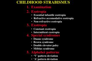
CLASSIFICATION OF STRABISMUS Strabismus can be classified in terms of: 1.DIRECTION OF DEVIATION- Hyper deviation- Hypo deviation- Divergent- Convergent
2. COMITANCY- concomitant / non-paralytic- incomitant / paralytic 3. CONSTANCY- constant- intermittent
CONCOMITANT(NON-PARALYTIC) • The movement of both eyes are full (there is no paresis) but only one eye is directed towards the fixated target. • The angle of deviation is constant and unrelated to the direction of gaze. • It is the common squint that is seen in childhood.
Under age of 6, it is rarely caused by serious neurological disease. It’s usually primary in this age group. • Strabismus arising later in life may have a specific and serious neurological basis.
Incomitant (paralytic) • The degree of misalignment varies with direction of the gaze. • One or more of the extraocular muscles or nerves may not be functioning properly, or normal movement may be restricted mechanically. • This type of strabismus may indicate either a nerve palsy or extraocular muscle disease.
Causes of isolated nerve palsies • Vascular disease..ex Dm, hypertension • Orbital disease…ex neoplasia • Trauma..most common cause of palsy of 4th,6th • Neoplasia… glioma • Raised intracranial pressure may cause a 3rd or 6th nerve palsy • Inflammation… sarcoidosis
Extraocular muscle disease • Dysthyroid eye disease • Myasthenia gravis • Ocular myositis • Ocular myopathy • Browns syndrome
Heterophoria • Heterophoria is a latent tendency for misalignment of the two eyes that becomes a manifest only if binocular vision is interrupted such as by covering one eye • A minor degree of heterophoria is normal for most individuals
Definations • Esotropia=inward movement • Exotropia=outward movement • Hypertropia=upward movement • Hypotropia=downward movement • prefix+phoria=(latent)the tendency to have squint when there is no concentration on a certain object….most common is exophoria
Strabismus testing • Corneal inspection • Hirschberg corneal light reflex test • Cover-uncover test
Corneal inspection • Have the patient look at the six cardinal positions of gaze to differentiate between concomitant and non-comitant
Hirschberg corneal light reflex • Objective assessment of ocular alignment • In newborn and often in young children it may be the only feasible method • Normally the light is reflected on each cornea symmetrically and in the same position relative to the pupil and visual axis on each side
In deviating eye the light reflection will be not centrally positioned and in direction opposite to that of the deviation • Example…pupil margin 15’,limbus 45’,iris 30’
Cover test • Easy, requires no special equipment and detect almost every case of tropia • Can be used in patients >6yrs • Have the patient look at the fixation point • Note which eye seems to be fixating • Cover it and observe the other • If it moves to pick up the fixation=>this eye was not directed toward the object of regard orginally • No shift on cover testing means there is no tropia
Cover test • Two types of cover test help to reveal a squint, especially if it is small and the examiner is unsure about the position of the corneal reflections. ● In the cover and uncover test, one eye is covered and the other eye is observed. If the uncovered eye moves to fix on the object there is a squint that is present all the time—a manifest squint. The test should then be carried out on the other eye. A problem arises when the vision in the squinting eye is reduced, and the eye may not be able to take up fixation. This emphasizes the need to test the vision of any patient with squint. If the cover and uncover test is normal (indicating no manifest squint) the alternate cover test should be done. ● In the alternate cover test, the occluder is moved to and fro between the eyes. If the eye that has been uncovered moves, then there is a latent squint.
History and exam • The patient complain of diplopia, there may be head posture to compensate for the eye to move in particular direction. • In third nerve palsy: • failure of adduction, elevation and depression of the eye. Ptosis in some cases, a dilated pupil due to involvement of autonomic fibres. • A fourth nerve palsy result in defective depression of eye when attempted in adduction. • A sixth nerve palsy results in faillure of abduction of the eye.
Management • Early detection • The most effective way to support fusion(binocular vision) is to treat amblyopia (failure of normal visual development) and equalize vision • Glasses can treat some or all of the esotropia In farsighted and may decrease deviation in near sided with myopia
Surgical corrections of misalignment may still be necessary for functional or cosmetic reasons • It must be stressed that surgery is not an alternative to glasses and patching when amblyopia • In paralytic strabimus treatment is directed to underlying pathology • Diplopia can be helped by fitting prisms to the patients glasses