Download
1 / 120
1.22k likes | 1.53k Views
Refractive Surgery Problem Solving. Dessi Fikaris, OD Jodi Abramson, MD TLC-White Plains, NY Special Thanks to Bill Tullo, OD December 16th 2010 2 hours. Dislodged flap Flap microstriae Epithelial Ingrowth DLK Loss of BCVA. Surface Ablation re-epithelialization Infectious Keratitis

E N D
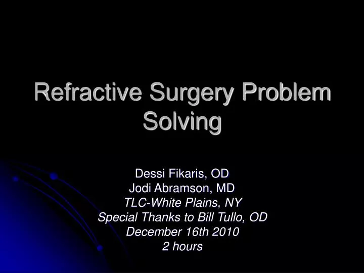
Refractive Surgery Problem Solving Dessi Fikaris, OD Jodi Abramson, MD TLC-White Plains, NY Special Thanks to Bill Tullo, OD December 16th 2010 2 hours
Dislodged flap Flap microstriae Epithelial Ingrowth DLK Loss of BCVA Surface Ablation re-epithelialization Infectious Keratitis Stromal Haze Dry Eye Enhancements Post-operative Considerations
RS • 31 year old male • 12 hours S/P uneventful LASIK OU • Patient phones with complaints of discomfort OU
Patient say’s My right eye became very uncomfortable about an hour after I got home and the vision is much better currently in my left eye.
When should RB be examined? Immediately!
RB’s Diagnosis #1 Slipped/Dislodged Flap
What would you recommend for RB? Return to surgeon and Lift and smooth flap
CLINICAL TESTS History UCVA OD and OS Slit lamp biomicroscopy Instructions RTO 3-5 days CLINICAL FINDINGS Dislodged flap* Flap Striae* DLK “SOS” Infection* SPK Poor UCVA LASIK Day One Pearls
AB • 25 year old female • 5 days S/P bilateral LASIK • Painless reduced VA in left eye since surgery
What tests would you perform on AB at 3-5 day PO visit? • UCVA OD and OS • Refraction and BCVA OD and OS • Slit lamp biomicroscopy • Tonometry • Dilate pupil • NaFl instillation
CLINICAL TESTS History UCVA Dry Rx BCVA (if not UCVA 20/20) Slit lamp biomicroscopy with NaFl Instructions, RTO 3 weeks CLINICAL FINDINGS Flap Striae DLK Infection Epithelial ingrowth SPK Refractive error Loss of BCVA 3-5 day Pearls - Critical Timing Return to Center Call docs
FLAP STRIAE 0.8 -0.3%1 Easier to see in Retro against a dilated pupil 1. Solomon R, Donnenfeld ED, Perry HD, Doshi S, Biser S Slitlamp stretching of the corneal flap after laser in situ keratomileusis to reduce corneal striae.J Cataract Refract Surg. 2003 Jul;29(7):1292-6
Retro-illumination… the key to detection
Flap Microstriae A. Often not visible at 1-day check B. Onset 24- 72 hours C. Will NOT resolve without treatment D. Common with high myopia E. Common with deep ablations F. Usually find small amounts of mixed astigmatism G. Common to report night vision complaints H. Rare with Intralase flaps
Striae • Only matters if Loss BCVA orsubjective quality of vision loss glare or halos
Treatment for flap microstriae 1. Caro ball or Q-tip smoothing 2. Flap Lift and Stretch 3. Flap Lift with Epithelial Debridement and hypotonic saline 4. Flap Suture 5. Therapeutic PTK
TS • 42 year old male • Right eye is sore to the touch since LASIK enhancement 1 month ago • Vision has declined in the right eye over past week
What tests would you perform on TS at 1 month PO visit? • UCVA in OD and OS • Refraction and BCVA in OD and OS • Slit lamp biomicroscopy OU • NaFl instillation OU • Tonometry OU • Corneal topography OU • Wavefront Aberrometry
CLINICAL TESTS History UCVA Dry Rx BCVA Slit lamp biomicroscopy with NaFl Instructions, RTO 2 months CLINICAL FINDINGS Flap Striae DLK “SOS” Epithelial ingrowth SPK Refractive error Loss of BCVA 1 Month Pearls - Critical Timing Return to surgical center Call surgical center
Most likely cause of discomfort in TS’s right eye # 3 - Epithelial ingrowth
Epithelial Ingrowth • Clinically significant risk with mechanical microkeratome = 2%1 Or 1:50 • Clinically significant risk with femtosecond microkeratome = 0.03%2 Or 1:3333 1. Asano-Kato N, Toda I, Hori-Komai Y, Takano Y, Tsubota K Epithelial ingrowth after laser in situ keratomileusis: clinical features and possible mechanisms. Am J Ophthalmol. 2002 Dec;134(6):801-7 2. Kamburoğlu G, Ertan A. Epithelial ingrowth after femtosecond laser-assisted in situ keratomileusis Cornea. 2008 Dec;27(10):1122-5
What are good reasons to treat Epitheilial ingrowth? • Epithelial cells within pupil with decreased BCVA • Persistent flap edge staining with NaFl • Progressive refraction or topographic changes • Flap melt • Persistent sore eye • Day time glare symptoms
The majority of Epithelial Ingrowth does not require treatment
CC 40 year old female S/P bilateral LASIK x 1 week Presents to your office for routine 1week post-op check up No symptoms reported Slitlamp biomicroscopy reveals “cloudy haze in right cornea”
Diagnosis for CC? #4 - Diffuse Lamellar Keratitis Incidence 0.18%1 1:562 1. Schallhorn SC, Venter JA. One-month outcomes of wavefront-guided LASIK for low to moderate myopia with the VISX STAR S4 laser in 32,569 eyes. J Refract Surg. 2009 Jul;25(7 Suppl):S634-41.
DLK may present • Focal area within interface • Diffuse throughout stromal interface • In an arcuate pattern • Stromal interface underneath the upper lid • Years after LASIK surgery
Mild –moderate DLK is associated with • No anterior chamber inflammation • No conjunctival injection • No Na Fl staining • No pain • Excellent prognosis!
Possible etiology of DLK includes A. Reaction to Staph. Endotoxins B. Contaminated sterilizer reservoir C. Excessive corneal manipulation D. Poor manufactured blades E. Mold or fungal contamination F. Excessive Intralase energy G. Unknown
Examination of CC reveals • Normal UCVA and BCVA • Na Fl reveals no staining • Applanation tonometry is normal
CC’s Treatment Plan Increase topical steroid q1h
How would you taper steroids? Topical Pred forte 1% q1h then taper slowly as biomicroscopy appearance improves
What if patient returns in 2 weeks and…? • Applanation tonometry measured 35 mmHg
CC’s Diagnosis DLK with ocular hypertension secondary to topical steroid
How does this change CC’s Treatment Plan? Add topical Beta Blocker bid
2 weeks post DLK Diagnosis • Applanation tonometry is 14 mmHg • Na Fl staining is negative • Vision is getting worse NOT better • Patient is taking topical Pred forte 1% q1h as prescribed
What is the MOST important additional test would you perform? Digital tonometry
CC’s Final Diagnosis DLK with fluid in the stromal interface
Delayed or mistreated DLK can result in all of the following • Stromal haze or scarring • Stromal flap melt • Flap microstriae • Uncontrolled glaucoma • Loss of BCVA
Flap microstriae Epithelial ingrowth Decentered ablations Central Islands Stromal Haze Irregular epithelium Irregular astigmatism High Order Aberrations Dry Eye Syndrome #5 – Loss of BCVA
Loss of BCVA • At 3-5 day PO • BCVA should be = to Pre-op BCVA • RGP Test if no obvious cause
Decentered Ablation with Induced Vertical Coma • 53 yo female presents in 2001 with complaints of glare, multiplopia, and “smeared vision” left eye • s/p LASIK for -8.0 D spherical myopia. • -1.75 -1.25 x 90 20/25. • Topo shows superior decentered ablation. • Patient undergoes wavefront-guided enhancement.
3 month post-op exam • Pleased with quality of vision • MR -.50 sphere 20/20 (vs. 20/25) • Wavefront refraction -0.66 -0.49 x 101 • Vertical coma -0.48 (vs. 1.39) • Topo: OZ nearly centered
Poor Vision after PRK 1995 • OD -6.25 -1.00 x 120, OS -6.75 -1.00 x 55 • PRK OU 1995. Ablation Zone 4.5 mm • OD: -1.75-1.50x 165, 20/30 • OS: -1.00 -1.25 x 25, 20/30 • Scotopic pupils 7 mm • Pachymetry: 490 OD, 505 OS • Post-op: c/o poor quality vision & glare OU • Dx: Small OZ OU • Tx: Custom Wavefront Guided PRK