Download
1 / 40
600 likes | 1.99k Views
BLOOD PRESSURE. Dr. Mullasari Ajit S. Director – Cardiology Madras Medical Mission. DEFINITION. The arterial BP, a measure of lateral force per unit area of vascular wall, is quantitated as millimeters of mercury (mmHg) or dynes per square centimeter (d/cm 2 ). HISTORY.

E N D
BLOOD PRESSURE Dr. Mullasari Ajit S. Director – Cardiology Madras Medical Mission
DEFINITION • The arterial BP, a measure of lateral force per unit area of vascular wall, is quantitated as millimeters of mercury (mmHg) or dynes per square centimeter (d/cm2).
HISTORY • 19TH Century- Jules Herisson –Sphygmograph • 1905, Dr. Nikolai Korotkoff (1874-1920), a Russian surgeon -a simple and precise technique to measure arterial pressure. • Korotkoff described four distinct phases of sounds: He failed to notice only the muffled second sound, which was demonstrated a little later.
Factors responsible for the peak systolic BP: 1. Volume and velocity of LV ejection, 2. The peripheral arteriolar resistance, 3. The distensibility of the arterial wall, 4.The viscosity of the blood, and 5. The end-diastolic volume in the arterial system
Factors responsible for the diastolic BP: 1. Blood viscosity, 2. Arterial distensibility, 3. Peripheral resistance to flow, and 4. Length of the cardiac cycle
Measurement of blood pressure: • 1.Direct methods 2.Indirect methods
Diretc method: • Use the electromanometer, a transducer that converts mechanical energy into an electric signal. • The artery is cannulated with a saline-filled catheter or needle that mechanically couples the circulation to the arterial manometer. • Pressures are recorded using atmospheric pressure as the zero reference level, and intravascular pressures are further referenced to the level of the heart by addition or subtraction of a gravitation factor
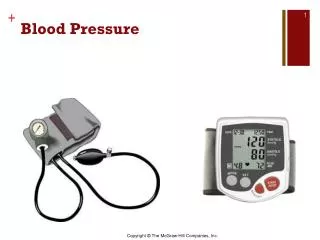
Indirect methods: • For the noninvasive evaluation of arterial BP, a pneumatic cuff with a mercury or aneroid manometer is the most frequently used technique.
Important Aspects of Blood Pressure measurement • Patient should sit or lie quietly for at least 5 minutes before the measurement is taken. • Seated comfortably, back supported, bared upper arm, legs uncrossed • Arm should be at heart level - the mid-point of the sternum • Cuff length/width - 80%/40% of arm circumference • Cuff should be deflated at <3 mm Hg per second • Column or dial should be read to nearest 2 mm Hg • First audible Korotkoff sound is systolic pressure; last sound is diastolic pressure • No talking between subject and observer
Measurements made while the patient is on an examining table do not fulfill these criteria and should preferably be made while the patient is seated in a chair. • Factors causing significant deviations in measured blood pressure: Room temperature, exercise, alcohol or nicotine consumption, positioning of the arm, muscle tension, bladder distension, talking, and background noise.
Choice of Blood Pressure Measurement Devices: The “gold standard” device for office blood pressure measurement has been the mercury sphygmomanometer. Cuff size: An undersized cuff - falsely high readings, and An oversized cuff - falsely low readings The recommended cuff sizes are:for arm circumference • 22 to 26 cm, 'small adult' cuff, 12 x 22 cm • 27 to 34 cm, 'adult' cuff: 16 x 30 cm • 35 to 44 cm, 'large adult' cuff: 16 x 36 cm • 45 to 52 cm, 'adult thigh' cuff; 16 x 42
Cuff Placement and Stethoscope: • First palpate the brachial artery • The lower end of the cuff should be 2 to 3 cm above the antecubital fossa to allow room for placement of the stethoscope • Pull the cuff snugly around the bare upper arm Phase 1 (systolic) and phase 5 (diastolic) Korotkoff sounds are best heard using the bell of the stethoscope over the palpated brachial artery in the antecubital fossa.
Traditionally, the sounds have been classified as 5 phases: • Phase I- appearance of clear tapping sounds corresponding to the appearance of a palpable pulse; • Phase II-sounds become softer and longer; • Phase III- sounds become crisper and louder; • Phase IV- sounds become muffled and softer; and Phase V- sounds disappear completely. The fifth phase is thus recorded as the last audible sound.
The sounds are thought to originate from a combination of turbulent blood flow and oscillations of the arterial wall. • Phase I corresponds to systolic pressure but underestimates the systolic pressure recorded by direct intra-arterial measurement. • Phase V corresponds to diastolic pressure but tends to occur before diastolic pressure determined by direct intra-arterial measurement. No clinical significance has been attached to phases II and III. • The Korotkoff sound method tends to give values for systolic pressure that are lower than the true intra-arterial pressure, and diastolic values that are higher
There has been disagreement in the past as to whether phase IV or V of the Korotkoff sounds should be used for recording diastolic pressure, but phase IV tends to be even higher than phase V when compared against the true intra-arterial diastolic pressure and is more difficult to identify than phase V. • There is now general consensus that the fifth phase should be used, except in situations in which the disappearance of sounds cannot reliably be determined because sounds are audible even after complete deflation of the cuff.
Use of phase IV as a diastolic pressure: • ·Pregnancy • ·Arteriovenous fistulas (eg, for hemodialysis), • ·Aortic insufficiency Most of the large-scale clinical trials that have evaluated the benefits of treating hypertension have used the fifth phase
Inflation/Deflation: • The cuff should initially be inflated to at least 30 mm Hg above the point at which the radial pulse disappears.(THIS AVOIDS THE AUSCUTATORY GAP) • The rate of deflation has a significant effect on blood pressure determination. • Deflation rates >2 mm per second can lead to a significant underestimation of systolic and overestimation of diastolic blood pressure. • It is recommended that a deflation rate of 2 to 3 mm Hg per second
Number of Measurements: • When a series of readings is taken, the first is typically the highest. • A minimum of 2 readings should be taken at intervals of at least 1 minute, and the average of those readings should be used to represent the patient’s blood pressure. • If there is >5 mm Hg difference between the first and second readings, additional (1 or 2) readings should be obtained, and then the average of these multiple readings is used.
Automated Methods When they are used in the office, the readings are typically lower than readings taken by a physician or nurse. Advantages : • ·Elimination of observer error, • ·Minimizing the white coat effect, and • ·Increasing the number of readings. Disadvantages: • The error inherent in the oscillometric method • The fact that epidemiologic data are mostly based on auscultated blood pressure measures
Required Competencies: • Vision- The observer must be able to see the dial of the manometer or the meniscus of the mercury column at eye level without straining or stretching, and must be able to read well enough to see the sphygmomanometer or digital display no further than 3 feet away. • Hearing- The observer must be able to hear the appearance and disappearance of Korotkoff sounds. • Eye/hand/ear coordination- This is required for the use of mercury and aneroid sphygmomanometers but not for the newer electronic technologies.
Effects of Body Position: • Blood pressure measurement is most commonly made in either the sitting or the supine position. • Sitting position- diastolic pressure is higher than when measured supine (by 5 mm Hg), although there is less agreement about systolic pressure. • Supine position: The systolic pressure is 8 mm Hg higher in the supine than the upright position. • If the back is not supported (as when the patient is seated on an examination table as opposed to a chair), the diastolic pressure may be increased by 6 mm Hg. • Crossing the legs: May raise systolic pressure by 2 to 8 mm Hg
In the supine position, the right atrium is approximately halfway between the bed and the level of the sternu; thus, if the arm is resting on the bed, it will be below heart level. • For this reason, when measurements are taken in the supine position the arm should be supported with a pillow. • In the sitting position, the right atrium level is the midpoint of the sternum or the fourth intercostal space
Effects of Arm Position: • If the upper arm is below the level of the right atrium- High BP • If the upper arm is above the level of the right atrium- low BP • Difference: 2 mm Hg for every inch above or below the heart level • These differences can be attributed to the effects of hydrostatic pressure
Differences Between the 2 Arms: • Blood pressure should be measured in both arms either in rapid succession or simultaneously; normally the measurements should differ by <10 mm Hg, independent of handedness. • As many as 20 percent of normal subjects, however, have a >10 mm Hg arm blood pressure difference.
Differences Between arms and legs: Systolic leg pressures may be as much as 20 mm Hg higher than arm pressures. • Greater leg-arm pressure differences are seen in patients with- • Severe AR (Hill sign) and • Patients with extensive and calcified lower peripheral arterial disease (PAD) • Aortic dissection • Coarctation of aorta • When there is a consistent interarm difference, the arm with the higher pressure should be used.
Blood Pressure Recording in Special Situations: • Elderly Patients:Blood pressure should also be taken in the standing position routinely because the elderly may have postural hypotension. • Arrhythmias:The blood pressure should be measured several times and the average value used. If severe regular bradycardia is present (eg, 40 to 50 bpm), deflation should be slower than usual to prevent underestimation of systolic and overestimation of diastolic blood pressure • Obese Patients:A longer and wider cuff is needed for adequate compression of the brachial artery in the obese patient with a very large upper arm.
Children: • Blood pressure is most conveniently measured in children by auscultation with a standard mercury sphygmomanometer. • Newborn–premature infants: a cuff size of 4-8 cm • Infants: 6-12 cm; and • Older children: 9-18 cm.
Isolated Systolic Hypertension: When the average systolic blood pressure is >140 and diastolic blood pressure is <90, the patient is classified as having isolated systolic hypertension. • Isolated Diastolic Hypertension: More commonly seen in some younger adults, isolated diastolic hypertension is defined as a systolic pressure <140 and a diastolic >90.
White-Coat Hypertension or Isolated Office Hypertension: • In 15% to 20% of people with stage 1 hypertension, blood pressure may only be elevated persistently in the presence of a health care worker, particularly a physician. • When measured elsewhere, including while at work, the blood pressure is not elevated. When this phenomenon is detected in patients not taking medications, it is referred to as white-coat hypertension (WCH). • The The commonly used definition is a persistently elevated average office blood pressure of >140/90 and an average awake ambulatory reading of <135/85 mm Hg. • In some patients, WCH may progress to definite sustained hypertension, and all need to be followed-up indefinitely with office and out-of-office measurements of blood pressure.
Masked Hypertension or Isolated Ambulatory Hypertension: • Somewhat less frequent than WCH but more problematic to detect is the converse condition of normal blood pressure in the office and elevated blood pressures elsewhere, eg, at work or at home. Lifestyle can contribute to this, eg, alcohol, tobacco, caffeine consumption, and physical activity away
Pseudohypertension: • When the peripheral muscular arteries become very rigid from advanced (often calcified) arteriosclerosis, the cuff has to be at a higher pressure to compress them. • The brachial or radial artery may be palpated distal to the fully inflated cuff in these instances (positive Osler sign). • When suspected, an intra-arterial radial artery blood pressure can be obtained for verification. • It was present in 7.2% of 3387 persons older than 59 years screened for the Systolic Hypertension in the Elderly Program (SHEP) study.
Orthostatic or Postural Hypotension: • Orthostatic hypotension is defined as a reduction of systolic blood pressure of at least 20 mm Hg or 10 mm Hg in diastolic blood pressure within 3 minutes of quiet standing. • An alternative method is to detect a similar fall during head-up tilt at 60 degrees
Coanda effect: • In supravalvular aortic stenosis the direction of the jet of flow tends to be directly directed into the innominate artery. This often results in the direct impact pressure of the central jet being transmitted to the right arm, thus making the right arm pressure higher than the left due to a Coanda effect
The Coanda Effect has been discovered in1930 by the Romanian aerodynamicist Henri-Marie Coanda (1885-1972). He had observed that a stream of air (or a other fluid) emerging from a nozzle tends to follow a nearby curved surface, if the curvature of the surface or angle the surface makes with the stream is not too sharp. • The Coanda Effect • In valvular aortic stenosis, the velocity of the jet is quicklydissipated beyond the stenotic orifice, preventing any sustainedhigh-velocity stream. • However, the smooth, annular narrowingof SVAS creates a "step" between the orifice and the ascendingaortic wall which enhances the natural affinity of a jet fora boundary wall and conserves the kinetic energy of the jetstream. In most patients with SVAS, the high-velocity streamis along the right aortic wall, causing disproportionately highpressure in the right arm.
A.Valvular aortic stenosis with mild poststenotic dilatation. Note abrupt transition between stenotic valve and ascending aortic wall. B. Supravalvular aortic stenosis. Note gradual transition (step) between annular stenosis and ascending aortic wall.
The Ankle-Brachial Index • The ankle-brachial index (ABI) is the ratio of the systolic blood pressure at the ankle divided by the higher of the two arm systolic blood pressures. • It reflects the degree of lower-extremity arterial occlusive disease, which is manifest by reduced blood pressure distal to stenotic lesions. Either the posterior tibial or dorsalis pedis artery pressures can be used. • The ABI is inversely related to disease severity. • A resting ABI <0.9 is considered abnormal. • An ABI <0.3 is consistent with critical ischemia, rest pain, and tissue loss. • For screening purposes, ABI is usually measured only at rest. However, a resting ABI >0.9 does not exclude significant PAD
Pulsus Paradoxus: • A normal person can exhibit a 10 mmHg drop in systolic pressure during normal inspiration. • A greater decline can be identified in patients with acute cardiac tamponade, constrictive pericarditis, severe obstructive lung disease, and restrictive cardiomyopathy. • Pulsus paradoxus is best detected by inflating the BP cuff above systolic pressure and then slowly releasing it. As the cuff pressure is gradually reduced, the BP sounds become audible during expiration. • The difference in pressure between the first audible sound heard on expiration and the pressure level at which the sounds are heard during all phases of respiration gives a measurement of magnitude of pulsus paradoxus.