Download
1 / 43
460 likes | 654 Views
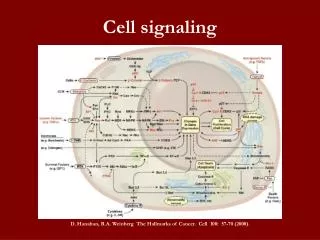
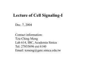
Lecture of Cell Signaling-I. Dec. 7, 2004. Contact information: Tzu-Ching Meng Lab 614, IBC, Academia Sinica Tel: 27855696 ext 6140 Email: tcmeng@gate.sinica.edu.tw. Phosphorylation is reversible. PTPs. P. P. P. P. Y. Y. Y. Y. Protein. Protein. Y. Y. P. P. PTKs.

E N D
Lecture of Cell Signaling-I Dec. 7, 2004 Contact information: Tzu-Ching Meng Lab 614, IBC, Academia Sinica Tel: 27855696 ext 6140 Email: tcmeng@gate.sinica.edu.tw
Phosphorylation is reversible PTPs P P P P Y Y Y Y Protein Protein Y Y P P PTKs
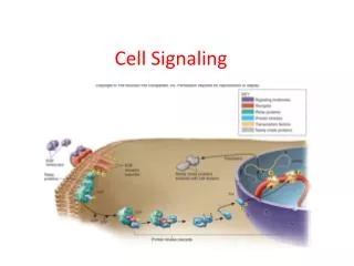
Protein modules in the control of intracellular signaling pathways Docking proteins function as platforms for the recruitment of signaling molecules
Models for activation of Signaling proteins A). By membrane translocation B). By conformational change C). By tyrosine phosphorylation
Signaling pathways activated by receptor tyrosine kinases Mechanisms for attenuation of receptor tyrosine kinases
Classification of human cytoplasmic protine tyrosine kinases
Activation of receptor tyrosine kinases Juxtamembrane region N-terminal kinase lobe Substrate precluding loop Substrate accessible loop C-terminal tail
Activation of c-Src • Two modes of intrinsic inhibition • by interactions between: • SH2 domain and • phosphorylated Y527; • (2) SH3 domain and • Polyproline region.
Activation of PKB/Akt PH domain precludes Kinase access by PDK-1
* *
* *
In most cases of CML, the leukemic cells share a chromosome abnormality not found in any nonleukemic white blood cells, nor in any other cells of the patient's body. This abnormality is a reciprocal translocation between one chromosome 9 and one chromosome 22. This translocation is designated t(9;22). It results in one chromosome 9 longer than normal and one chromosome 22 shorter than normal. The latter is called the Philadelphia chromosome and designated Ph1. Expression of a fusion PTK p210 Brc-Abl
W W W W FN FN FN FN FN FN FN FN FN FERM SH2 SH2 SH2 W FERM The Protein Tyrosine Phosphatase Superfamily (HCx5R) ‘Classical’ pTyr Specific PTPs (HCSAGxGRxG) Dual Specificity Phosphatases (HCxxGxxR) PTEN Non-transmembrane PTPs Receptor-type PTPs VHR-like Cdc25 FN FN FN FN FN FN FN MAM FN FN FN C2 FN FN FN FN FN FN FN FN FN FN FN FN FN FYVE VHR VH1 MKP-1 MKP-2 MKP-3 MKP-4 MKP-5 KAP (Cdi1) FYVE- DSP Cdc25A Cdc25B Cdc25C PTEN (MMAC1) P E S T PTPb DEP1 SAP1 GLEPP1 PTPH1 MEG1 PTPD1 PTPD2 PTPBAS MEG2 SHP1 SHP2 CD45 PTP1B TCPTP PEST LyPTP Heavily glycosylated PTPm PTPk PTPr PTPl LAR PTPs PTPd PTPa PTPe PTPg PTPz FERM domain PTP domain Carbonic anhydrase-like Fibronectin III Like repeat Src Homology domain 2 FN Cadherin-like DSP domain Retinaldehyde Binding protein-like Merpin/A5/m domain MAM FYVE-domain FYVE PEST-like PEST Lipid binding domain Immuno- globulin-like C2 PDZ domain Tonks NK & Neel BG, Curr Opin Cell Biol. 2001, 13(2):182-95
Classification of Protein Tyrosine Phosphatases Non-transmembrane PTPs Receptor-like PTPs Andersen et al., Mol Cell Biol, 21, 7117, 2001
C-terminal - ER targeting - Proteolytic cleavage Proline rich segment - SH3 binding sites Alternative splicing - Nucleus vs Cytoplasmic FERM domain - Subcellular targeting (e.g. cytoskeletal proteins) PDZ domain(s) - Protein-Protein interactions SH2 domains - Plasma membrane signaling complexes - Auto-inhibition PEST domain - Protein-Protein Interactions BRO1 domain - Functionally uncharacterised; (Found in a number of signal transduction proteins) - Vesicle associated His-domain - Functionally uncharacterised • Cellular retinaldehyde • binding protein-like • - Golgi targeting • - Secretory vesicles • - Putative lipid-binding domain Functional Diversity Through Targeting and Regulatory Domains
Location of conserved motifs in 3D IVMxT (M6) KCxxYWP (M7) WPDxGxP (M8) TxxD FWxMxW (M5) QTxx QYxF (M10) PxxV HCSAGxGRTG (M9) IAxQGP (M4) DxxRVxL (M2) NxxKNRY (M1) DYINA (M3) http://ptp.cshl.edu
Conserved fold of PTP domains N-terminal Central a3-helix Andersen et al Mol. Cell. Biol. 2001
Protein Tyrosine Phosphatase 1B WPD loop
W W W W FN FN FN FN FN FN FN FN FN FERM SH2 SH2 SH2 W FERM The Protein Tyrosine Phosphatase Superfamily (HCx5R) ‘Classical’ pTyr Specific PTPs (HCSAGxGRxG) Dual Specificity Phosphatases (HCxxGxxR) PTEN Non-transmembrane PTPs Receptor-type PTPs VHR-like Cdc25 FN FN FN FN FN FN FN MAM FN FN FN C2 FN FN FN FN FN FN FN FN FN FN FN FN FN FYVE VHR VH1 MKP-1 MKP-2 MKP-3 MKP-4 MKP-5 KAP (Cdi1) FYVE- DSP Cdc25A Cdc25B Cdc25C PTEN (MMAC1) P E S T PTPb DEP1 SAP1 GLEPP1 PTPH1 MEG1 PTPD1 PTPD2 PTPBAS MEG2 SHP1 SHP2 CD45 PTP1B TCPTP PEST LyPTP Heavily glycosylated PTPm PTPk PTPr PTPl LAR PTPs PTPd PTPa PTPe PTPg PTPz FERM domain PTP domain Carbonic anhydrase-like Fibronectin III Like repeat Src Homology domain 2 FN Cadherin-like DSP domain Retinaldehyde Binding protein-like Merpin/A5/m domain MAM FYVE-domain FYVE PEST-like PEST Lipid binding domain Immuno- globulin-like C2 PDZ domain Tonks NK & Neel BG, Curr Opin Cell Biol. 2001, 13(2):182-95
Sequence alignment of amino acid residues at phosphatase motif among human DSPs
MAPK and SAPK pathway in mammalian cells T-x-Y at the activation loop
Inactivation of MAP kinases (ERK) by threonine or tyrosine dephosphorylation
PTPs and Cancer Refinement of PTP chromosomal positions allows for genetic disease linkage studies 19 PTP chromosomal regions are frequently deleted in human cancers 3 PTP chromosomal regions are frequently duplicated in human cancers
PTPs and Cancer PTEN Tumor Suppressor Mutated in various human cancers. Cowden disease DEP1 Tumor suppressor Colon cancer susceptibility locus Scc1 (QTL in mice) PTPkTumor Suppressor Primary CNS lymphomas SHP2 Noonan Syndrome Developmental disorder affecting 1:2500 newborn Stomach Ulcers Target of Helicobacter pylori Cdc25 Cell Cycle Control Target of Myc and overexpressed in primary breast cancer PRL-3 Metastasis Upregulated in metastases of colon cancer FAP-1 Apoptosis Upregulated in cancers, inhibits CD95-mediated apoptosis
PTPs as Drug Targets Immunosupression Diabetes & Obesity Autoimmunity & Allergy PTPs Infectious diseases Cancer Epilepsy
S (Inactive) P P PTK PTK (Inactive) (Active) S P P (Active) Interactions Between PTKs and PTP– (1) PTPs function as NEGATIVE Regulators of Signal Transduction Autophosphorylation PTP PTP
P P Interactions Between PTKs and PTPs—(2) PTPs function as POSITIVE Regulators of Signal Transduction PTP S S (Inactive) (Active) PTK
Important references • Hunter, T. (2000) Signaling-2000 and beyond. Cell, 100: 113-127 • J. Schlessinger (2000) Cell signaling by receptor tyrosine kinases. • Cell, 103: 211-225 • 3. Myers, M. et al. (2001) TYK2 and JAK2 are substrates of protein • tyrosine phosphatase 1B. J. Biol. Chem., 276: 47771-47774 • Andersen, J. N. et al. (2001) Structural and evolutional relationships • among protein tyrosine phosphatase domains. Mol. Cell. Biol., • 21: 7117-7136 • 5. Tonks, N. K. (2003) PTP1B: From the sidelines to the front lines. • FEBS Letters, 546: 140-148
Additional references • Blume-Jensen, P. Hunter, T. (2000) Oncogenic kinase signaling. • Cell, 100: 113-127. • 2. Palka, H., Park, M. and Tonks, N.K. (2003) Hepatocyte growth factor • receptor kinase Met is a substrate of the receptor protein tyrosine • phosphatase DEP-1. J. Biol. Chem., 278: 5728-5735. • 3. Salmeen, A. et al. (2000) Molecular basis for the dephosphorylation • of the activation segment of the insulin receptor by protein tyrosine • phosphatase 1B. Mol. Cell, 6: 1404-1412. • 4. Meng, T.C. et al (2004) Regulation of insulin signaling through • reversible oxidation of the protein-tyrosine phosphatases TC45 and • PTP1B. J. Biol. Chem., 279: 37716-37725.