Download
1 / 25
250 likes | 357 Views
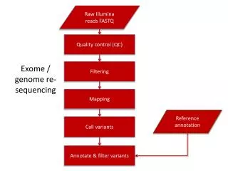
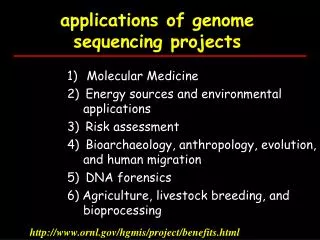
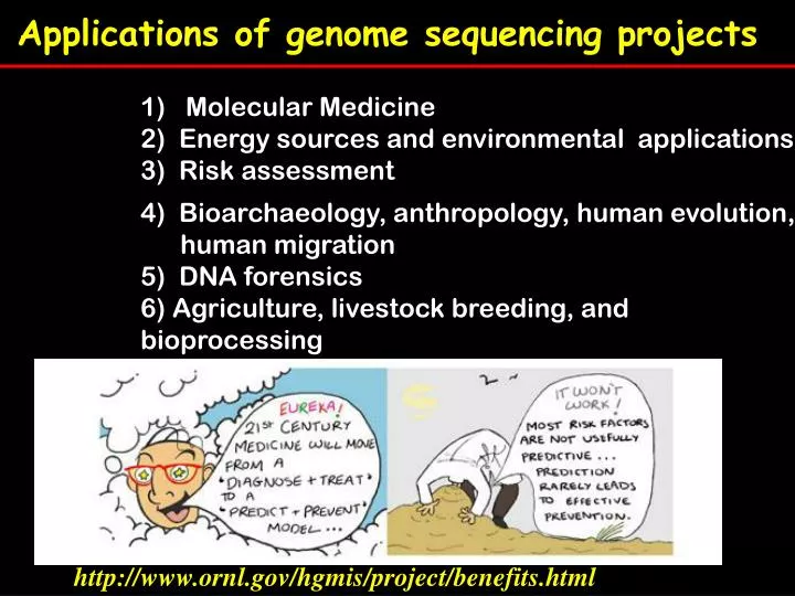
Applications of genome sequencing projects. 1) Molecular Medicine 2) Energy sources and environmental applications 3) Risk assessment. 4) Bioarchaeology, anthropology, human evolution, human migration 5) DNA forensics 6) Agriculture, livestock breeding, and bioprocessing.

E N D
Applications of genome sequencing projects • 1) Molecular Medicine • 2) Energy sources and environmental applications • 3) Risk assessment • 4) Bioarchaeology, anthropology, human evolution, human migration • 5) DNA forensics • 6) Agriculture, livestock breeding, and bioprocessing http://www.ornl.gov/hgmis/project/benefits.html
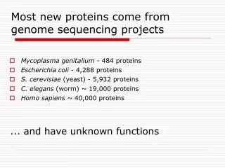
Molecular medicine improved diagnosis of disease eearlier detection of genetic predisposition to disease rational drug design gene therapy and control systems for drugs ppharmacogenomics "custom drugs"
The spectrum of human diseases Cystic fibrosis thalassemia Huntington’s <5% cancer
‘Mendelian’ diseases (<5%) Autosomal dominant inheritance: e.g huntington’s disease Autosomal codominant inheritance e.g Hb-S sickle cell disease Autosomal recessive inheritance: e.g cystic fibrosis, a & b thalassemias X-linked inheritance: e.g Duchenne muscular dystrophy (DMD)
How to identify disease genes • Identify pathology • Find families in which the disease is segregating • Find ‘candidate gene’ • Screen for mutations in segregating families
How to map candidate genes 2 broad strategies have been used A. Position independent approach (based on knowledge of gene function) 1)biochemical approach 2) animal model approach B. Position dependent approach (based on mapped position)
Position independent approach 1) Biochemical approach: when the disease protein is known E.g. Factor VIII haemophilia Blood-clotting cascade in which vessel damage causes a cascade of inactive factors to be converted to active factors
Blood tests determine if active form of each factor in the cascade is present Fig. 11.16 c
Techniques used to purify Factor VIII and clone the gene Fig. 11.16 d Fig. 11.16 d Hartwell
2) Animal model approach compares animal mutant models for a phenotypically similar human disease. E.g. Identification of the SOX10 gene in human Waardenburg syndrome4 (WS4) Dom (dominant megacolon) mutant mice shared phenotypic traits similar to human patient with WS4 (Hirschsprung disease, hearing loss, pigment abnormalities) WS4 patients screened for SOX10 mutations confirmed the role of this gene in WS4. Waardenburg Dom mouse Hirschsprung
B) Positional dependent approach Positional cloning identifies a disease gene based on only approximate chromosomal location. It is used when nature of gene product / candidate genes is unknown. Candidate genes can be identified by a combination of their map position and expression, function or homology
B) Positional Cloning Steps Step 1 – Collect a large number of affected families as possible Step 2- Identify a candidate region based on genetic mapping (~ 10Mb or more) Step 3- Establish a transcript map, cataloguing all the genes in the region Step 4- Identify potential candidate genes Step 5 – confirm a candidate gene
Step 2- Identifying a candidate region Genetic map of <1Mb Genetic markers: RFLPs, SSLPs, SNPs Lod scores: log of the odds: ratio of the odds that 2 loci are linked or not linked need a lod of 3 to prove linkage and a lod of -2 against linkage Halpotype maps HapMap published in Oct27 2005 Nature
Step 3– transcript map which defines all genes within the candidate region Search browsers e.g. Ensembl Computational analysis • Usually about 17 genes per 1000 kb fragment • Identify coding regions, conserved sequences between species, exon-like sequences by looking for codon usage, ORFs, and splice sites etc Experimental checks – double check sequences, clones, alignments etc Direct searches – cDNA library screen
Step 4 – identifying candidate genes Expression: Gene expression patterns can pinpoint candidate genes • RNA expression by Northern blot or RT-PCR or microarrays • Look for misexpression (no expression, underexpression, overexpression) CFTR gene Northern blot analysis reveals only one of candidate genes is expressed in lungs and pancreas
Step 4 – identifying candidate genes Function: Look for obvious function or most likely function based on sequence analysis e.g. retinitis pigmentosa Candidate gene RHO part of phototransduction pathway Linkage analysis mapped disease gene on 3q (close to RHO) Patient-specific mutations identified in a year
Step 4 – identifying candidate genes Homology: look for homolog (paralog or ortholog) Both mapped to 5q Marfan syndrome fibrillin gene FBN1 Beals syndrome fibrillin geneFBN2
Step 4 – identifying candidate genes Animal models: look for homologous genes in animal models especially mouse e.g. Waardenburg syndrome type 1 Linkage analysis localised WS1 to 2q Splotch mouse mutant showed similar phenotype Could sp and WS1 be orthologous genes? Pax-3 mapped to sp locus Homologous to HuP2 Splotch mouse WS type1
Step 5 – confirm a candidate gene • Mutation screening • Sequence differences • Missense mutations identified by sequencing coding region of candidate gene from normal and abnormal individuals • Transgenic model • Knockout / knockin the mutant gene into a model organism • Modification of phenotype
Transgenic analysis can prove candidate gene is disease locus Fig. 11.21
Figure 1 | Genetic and chemical-genetic approaches identify genes and proteins, respectively, that regulate biological processes. a | Forward genetics entails introducing random mutations into cells, screening mutant cells for a phenotype of interest and identifying mutated genes in affected cells. In the example shown, yeast cells are randomly mutated, cells showing a large-bud phenotype are selected, and genes mutated in these cells are identified. Reverse genetics entails introducing a mutation into a specific gene of interest and studying the phenotypic consequences of the mutation in a cellular or organismal context. In the example shown, a single mutated gene is introduced into yeast cells and a large-bud phenotype is observed. b | Forward chemical-genetics entails screening exogenous ligands in cells, selecting a ligand that induces a phenotype of interest, and identifying the protein target of this ligand. In the example shown, one compound that induces a large-bud phenotype is selected and the protein target of this ligand is subsequently identified. Reverse chemical-genetics entails overexpressing a protein of interest, screening for a ligand for the protein, and using the ligand to determine the phenotypic consequences of altering the function of this protein in a cellular context. In the example shown, a ligand for a specific protein is found to induce a large-bud phenotype.
Reading HMG3 by T Strachan & AP Read : Chapter 14 AND/OR Genetics by Hartwell (2e) chapter 11 Optional Reading on Molecular medicine Nature (May2004) Vol 429 Insight series • human genomics and medicine pp439 (editorial) • predicting disease using medicine by John Bell pp 453-456.