Download
1 / 77
1.06k likes | 2.81k Views
Goals. Learn about anatomy and development of the retina?What do all those stages and zones mean?"Understand the devastating visual consequences of ROPUnderstand your important role in the ROP teamShow lots of pictures to keep you awake. Definition of ROP. Abnormal retinal development in premature babiesSome of these infants will have an interruption of normal retinal growth followed by abnormal proliferation of blood vessels and fibrous tissue.

E N D
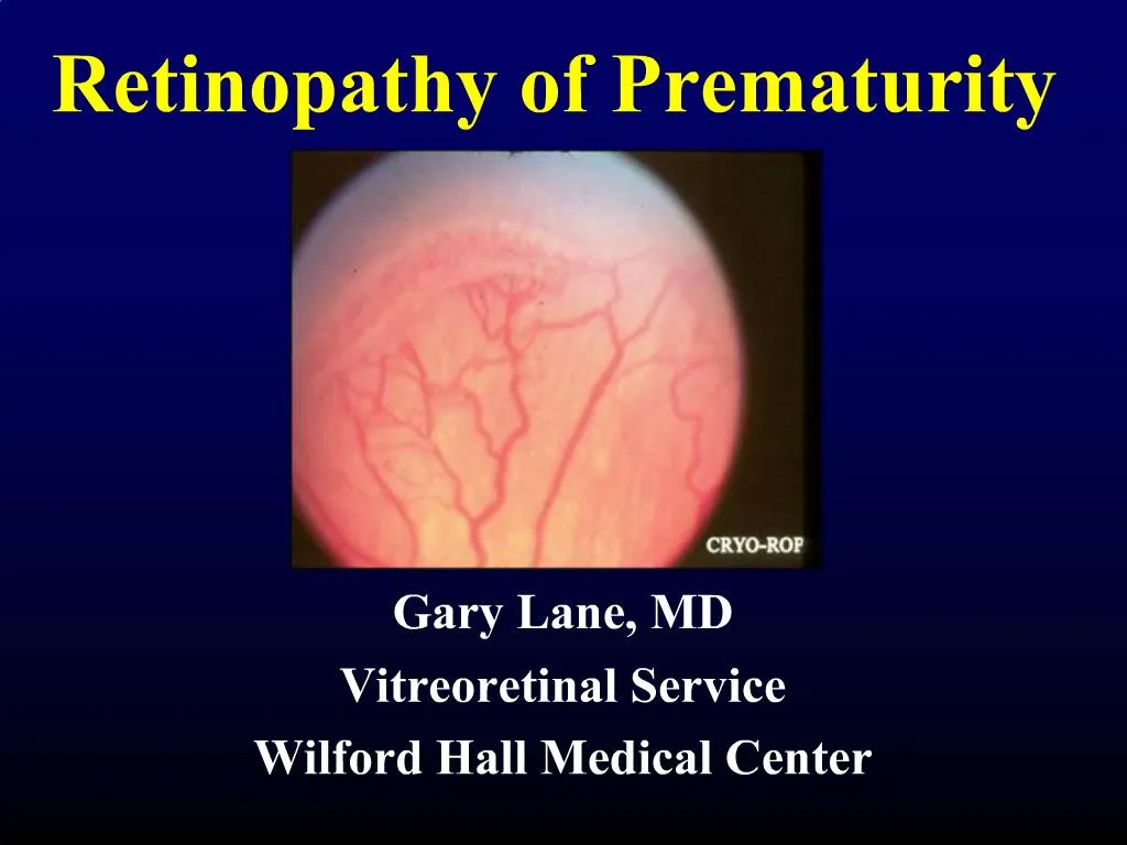
1. Retinopathy of Prematurity Gary Lane, MD
Vitreoretinal Service
Wilford Hall Medical Center
2. Goals Learn about anatomy and development of the retina
�What do all those stages and zones mean?�
Understand the devastating visual consequences of ROP
Understand your important role in the ROP team
Show lots of pictures to keep you awake
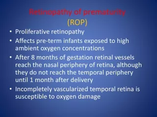
3. Definition of ROP Abnormal retinal development in premature babies
Some of these infants will have an interruption of normal retinal growth followed by abnormal proliferation of blood vessels and fibrous tissue
4. Who gets ROP? 66% of infants ? 1,250g develop some ROP
82% of infants ? 1,000g develop some ROP
9% become eligible for treatment
6. Retinal Anatomy 101
7. Normal vascular development
8. VEGF and IGF-1 Schematic representation of IGF-IyVEGF control of blood vessel development in ROP.
In utero, VEGF is found at the growing front of vessels. IGF-I is sufficient to allow vessel growth.
(B) With premature birth, IGF-I is not maintained at in utero levels and vascular growth ceases, despite the presence of VEGF at the growing front of vessels. Both endothelial cell survival (Akt) and proliferation (mitogen-activated protein kinase) pathways are compromised. With low IGF-I and cessation of vessel growth, a demarcation line forms at the vascular front. High oxygen exposure (as occurs in animal models and in some premature infants) may also suppress VEGF, further contributing to inhibition of vessel growth.
(C) As the premature infant matures, the developing but nonvascularized retina becomes hypoxic. VEGF increases in retina and vitreous. With maturation, the IGF-I level slowly increases.
(D) When the IGF-I level reaches a threshold at ~34 weeks gestation, with high VEGF levels in the vitreous, endothelial cell survival and proliferation driven by VEGF may proceed. Neovascularization ensues at the demarcation line, growing into the vitreous. If VEGF vitreal levels fall, normal retinal vessel growth can proceed. With normal vascular growth and blood flow, oxygen suppresses VEGF expression, so it will no longer be overproduced. If hypoxia (and elevated levels of VEGF) persists, further neovascularization and fibrosis leading to retinal detachment can occur.
Hellstrom et al. PNAS
Schematic representation of IGF-IyVEGF control of blood vessel development in ROP.
In utero, VEGF is found at the growing front of vessels. IGF-I is sufficient to allow vessel growth.
(B) With premature birth, IGF-I is not maintained at in utero levels and vascular growth ceases, despite the presence of VEGF at the growing front of vessels. Both endothelial cell survival (Akt) and proliferation (mitogen-activated protein kinase) pathways are compromised. With low IGF-I and cessation of vessel growth, a demarcation line forms at the vascular front. High oxygen exposure (as occurs in animal models and in some premature infants) may also suppress VEGF, further contributing to inhibition of vessel growth.
(C) As the premature infant matures, the developing but nonvascularized retina becomes hypoxic. VEGF increases in retina and vitreous. With maturation, the IGF-I level slowly increases.
(D) When the IGF-I level reaches a threshold at ~34 weeks gestation, with high VEGF levels in the vitreous, endothelial cell survival and proliferation driven by VEGF may proceed. Neovascularization ensues at the demarcation line, growing into the vitreous. If VEGF vitreal levels fall, normal retinal vessel growth can proceed. With normal vascular growth and blood flow, oxygen suppresses VEGF expression, so it will no longer be overproduced. If hypoxia (and elevated levels of VEGF) persists, further neovascularization and fibrosis leading to retinal detachment can occur.
Hellstrom et al. PNAS
11. ROP is caused by prematurity Oxygen is just one of several important factors
13. Retinopathy of Prematurity ICROP � Staging
Level of abnormal vascular response observed
17. Stage 3 ROP
21. Plus disease
24. QUIZ
26. Stage 4 (A-extramacular and B-macular)
27. Retinopathy of Prematurity ICROP � Staging
Stage 4
Subtotal retinal detachment
4a - extrafoveal
28. Retinopathy of Prematurity ICROP � Staging
Stage 4
Subtotal retinal detachment
4b � RD involving fovea
30. Retinopathy of Prematurity ICROP � Staging
Stage 5
Open funnel
Closed funnel
33. Stage 5 Total retinal detachment
�Retrolental fibroplasia�
34. Stage 5 ROP 25-50% anatomic success
� of these able to reach out and grab an object
Late onset (into adulthood) angle closure
Late onset RRD
15%, most requiring multiple surgeries
35. AP-ROP Aggressive Posterior ROP
A.K.A. Rush Disease
38. Screening At least 2 dilated exams for:
All infants weighing less than 1500 grams at birth or born prior to 32* weeks gestation
1st exam by age 31 weeks or 4 weeks after birth, whichever is later
Follow-up exams vary from every few days to every few weeks depending location (Zone) of ROP
Exams continue if treatment is needed and are discontinued if regression occurs
Selected infants between 1500-2000 grams with unstable clinical course
39. Threshold ROP: treatment
40. Early Treatment of Retinopathy of Prematurity 317 infants with bilateral high risk ROP
1 eye randomized to early treatment
RM-ROP2 is a risk analysis program based on natural history data from CRYO-ROP
Fellow eye treated at threshold per CRYO-ROP
41. The �New Threshold� Any Zone I ROP with Plus
Zone I ROP Stage 3 with or without Plus
Zone II Stage 2 or 3 with Plus
Pearl: Infants usually reach threshold at about their due date or 38 weeks gestational age
42. Retinopathy of Prematurity Treatment
Cryotherapy
43. Retinopathy of Prematurity Treatment
Laser photocoagulation
46. Laser vs. Cryo Depends on clear view
Minimal conj injection
~1000 spots
Cataract (ischemia)
Less destructive with comparable results
Useful in limited view
Massive chemosis
50-60 spots
Vitreous hemorrhage
Extensive peripheral damage
47. Lens sparing vitrectomy
48. Stage 5 ROP Remove lens
+/-Remove iris
+/- open sky approach
DO NOT create a break
Include a scleral buckle
Tamponade with healon
51. ROP and Oxygen PaO2 in utero is about 30mmHg
SpO2 of 70%
Repeated episodes of hypo and hyperoxia are thought to be important in the pathogenesis of ROP
Can manipulating PaO2 levels change the course of the disease?
From Iowa Neonatology Handbook
52. ROP and SpO2 Chow, et al. Pediatrics 2003
Implemented SpO2 guidelines
Careful titration to avoid hypoxia and hyperoxia
Target range of 85-93% for high risk babies
54. ROP and Oxygen Brain is similar to the retina
Periventricular leukomalacia is strongly correlated with sustained hyperoxia
Calls for large multi-center randomized controlled trials
Several trials in various countries will be pooled
BOOST 2
85-89% vs. 91-95%
55. ROP and Oxygen Provocative Results
Anecdotal evidence
Confounding variables
Needs a randomized, control trial
Caveat: Retinal surgeons don�t manage babies � you do
Consider risk of pulmonary disease
56. ROP: Creating a Safety Net Adapted from:
Retinopathy of Prematurity: Creating a Safety Net
Anne M. Menke, R.N., Ph.D.
OMIC Risk Manager
57. Case Study A premature infant was born at 29 weeks gestation and weighed 1200grams
Baby was examined and noted to have Stage 1 ROP, Zone 2, follow-up 4 weeks
58. Case Study Baby was transferred to another NICU
No further eye exams were performed
3 months later, pediatrician received the discharge summary from the first hospital and saw ROP mentioned
Child referred to ophthalmologist who diagnosed bilateral Stage V detachments
59. Safety Net Elements Clarification of physician roles
Risk analysis of current process of care
Hospital ROP Tracking System
Coordination of transfer of care
Ongoing education of ROP team: physicians, nurses and parents
60. Physician-Patient Relationship Screening infants in the NICU creates many difficulties
Once a physician assumes responsibility for a patient, he or she creates a physician-patient relationship from which certain legal duties arise
61. Physician-Patient Relationship Failure to provide follow-up can result in a claim of patient abandonment and harm from failure to follow-up.
Breakdowns in the follow-up process are the single most common cause of delays in the diagnosis and treatment of ROP.
62. Inpatient Physician Roles NICU Attending � primary care provider
responsible for coordinating all care until the baby is discharged from the hospital or transferred to another facility
Ophthalmologist � consultant
After examining the baby and determining a follow-up interval, care is finished unless another request is made
63. Outpatient Physician Roles In outpatient care, the ophthalmologist assumes primary responsibility for ROP follow-up until completion
Must follow-up on missed appointments and non-compliance
64. Risk Analysis of Current Process of Care Does every step have a person with assigned responsibility?
What are the key vulnerabilities?
Most ROP lawsuits occur when baby is lost to follow-up
What have been the past failures?
65. Hospital Tracking System Single person designated as ROP coordinator, with backup when on vacation
ROPC tracks baby until
Ophthalmologist determines that ROP exams are no longer necessary
Baby is discharged or transferred AND an ophthalmologist accepts care AND an appointment is scheduled
66. Key Vulnerability: Transfers and Discharges Hospital must:
Write a discharge order that documents future ROP care
ROP follow-up appointment is scheduled
Medical record and parent�s contact information have been forwarded
67. Education as a Safety Net Parents
NICU attendings
Nurses
Residents
68. Educating Parents & Caregivers Parents should be aware of the risk of blindness from untreated ROP, and this should be documented in the chart
Parents should read and sign �Caregivers: Read this about premature baby�s eyes�
69. Education of NICU team NICU attendings, residents and nurses need periodic education about ROP
Attitudes of nurses about ROP exams, positive or negative, are quickly communicated to the parents
70. ROP, Nursing and Parents ROP tends to get bad right around the due date
Usually after the baby has made through all the other tough times and is ready to go home
Sepsis, RDS, etc.
Abstract disease � difficult to understand
71. Message to Parents ROP is caused by prematurity
It�s not the fault of doctors, nurses or ventilators
Everyone is stressed out by ROP exams
�That�s gross�
ROP exams prevent blindness
Same anesthetic and lid speculum as for cataract surgery or LASIK
Parents need to follow up after discharge
72. ROP Follow-up No baby should go home without a car seat
And
No ROP baby should go home without a confirmed follow-up appointment in Ophthalmology
73. Review Zones refer to location
Zone 1 is most immature, worst prognosis
Stage refers to extent of disease
Stage 3 requires close observation or treatment
Stage 4 or 5 involve retinal detachments
Plus refers to presence of vascular shunting
Treatment is laser of peripheral retina
74. Review ROP tends to get bad right around due date
Most common cause of bad outcomes and lawsuits in ROP is missed follow-up appointments after hospital discharge or transfer
Good care of ROP babies requires education and a team approach between doctors, nurses and parents
76. No baby deserves to go blind from ROP
77. Questions
78. Www.ROPARD.com