Download
1 / 31
330 likes | 507 Views
Retinopathy of prematurity: Altered development. A disorder with a uniquely American heritage. Early History. Silverman, WA. Retrolental Fibroplasia: A Modern Parable. Monographs in Neonatology. 1980 Dr. Stewart Clifford, Boston pediatrician discovers first case -1941

E N D
Retinopathy of prematurity:Altered development A disorder with a uniquely American heritage
Early History • Silverman, WA. Retrolental Fibroplasia: A Modern Parable. Monographs in Neonatology. 1980 • Dr. Stewart Clifford, Boston pediatrician discovers first case -1941 • Dr. Harry Messenger, Boston ophthalmologist coined the term RLF
RLF National Cooperative Study Relative Risk = 3.6 ( 95% CI 1.7 – 7.75)
Normal oxygen values Avery et al. Neonatology: Pathophysiology and Management of the Newborn, 4th ed. Pg 130, table 11-2.
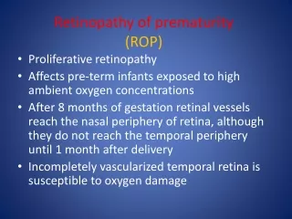
Retinal vascular development (ontogeny) • The choroidal vessels can supply the thin retina via diffusion • The retinal nerve cells (photoreceptors) develop from the optic nerve to the periphery • Additional blood supply develops as the retinal nerve cell layer becomes thicker
Ontogeny of the retinal vascular bed • Inner vascular plexus • Within the nerve fiber layer • Capillaries appear around the 16th week of gestation and reach the ora serrata at about 32 – 36 weeks gestation nasally and temporally just before term • Vasculogenesis
The goal – supply blood to the maturing retina http://www.tsbvi.edu/Outreach/seehear/winter98/ICROP.gif
Ontogeny of the retinal vascular bed • Outer vascular plexuses • Develops later in gestation and continues to develop postnatally • Capillaries arise as cellular buds from the innermost vessels • Angiogenesis
When ROP develops – How bad is it ? • Stage One – A line of demarcation between the vascular and avascular retina • Stage Two – The line comes a ridge • Stage Three – The ridge is associated with neonvascularization entering the vitreous
When ROP develops – How bad is it ? • Stage Four – Subtotal detachment of the retina • IV – A is extrafoveal detachment • IV – B the detachment includes the fovea • Stage Five – Total Detachment • The old retrolental fibroplasia An International Classification of Retinopathy of Prematurity. Arch Ophthalmol. 1987;105: 906-912.
When ROP develops – How bad is it ? • Plus Disease – very tortuous vessels implying high blood flow; bad • Rush Disease – Plus disease in zone 1
Stage One http://ropard.org/ The Association for Retinopathy of Prematurity and Related Diseases
ROP – A disease that can regress Pediatrics. 2005;116:15 – 23.
Incidence inversely proportional to gestational age at birth
Incidence inversely proportional to gestational age at birth
Prevention of severe disease • Primary – decrease the number of infants born at the gestations with highest risk • Secondary • An agent that will prevent the retinal blood vessel drop out after birth in very premature infants • Limit the vasoproliferative phase • Safe oxygen administration
Prevention of severe disease • Cryotherapy and laser therapy limit the vasoproliferative phase by destroying the avascular retina once THRESHOLD has been reached • Intravitreal bevacizumab (Avastin) injection
Prevention of severe diseaseCyrotherapy outcome at 5 ½ years Arch Opthalmol. 1996;224:417-424
Earlier treatment of disease in Zone One Arch Opthalmol. 2003; 121:1684-96
Limit excessive oxygen exposure • Conclusion: Inappropriate oxygen use is a neonatal health hazard associated with aging, DNA damage and cancer, retinopathy of prematurity, injury to the developing brain, infection and others. Neonatal exposure to pure O2, even if brief, or to pulse oximetry >95% when breathing supplemental O2 must be avoided as much as possible • Sola, A, et al. Acta Paediatrica. 96(6):801-812, June 2007.
Limit oxygen exposure Chow et al. Pediatrics. 2003;111:339-45
Screen • All infants with birth weights less than 1500 grams or gestational age less than 32 weeks • Begin at 4 to 6 weeks • Continue until mature (vascularized to the periphery)
Other ophthalmologic sequelae Cats B. and Tan K. J Ped Opthamal & Strabismus. 1989:271-75
Myopia related to ROP Quinn GE et al.Opthalmology 1998; 105:1292-1299
Myopia related to ROP Quinn GE et al.Opthalmology 1998; 105:1292-1299