Download
1 / 36
410 likes | 542 Views
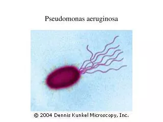
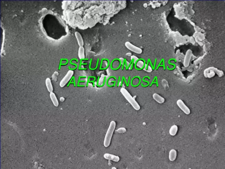
PSEUDOMONAS AERUGINOSA. Pseudomonas aeruginosa. Gram-negative bacillus long and slender bacilli Pigmented growth on agar and distinct odor usually green or red and smells like grapes or juicy fruit gum Ubiquitous in environment, especially in water

E N D
Pseudomonas aeruginosa • Gram-negative bacillus • long and slender bacilli • Pigmented growth on agar and distinct odor • usually green or red and smells like grapes or juicy fruit gum • Ubiquitous in environment, especially in water • Hospital environment loaded with this pathogen • Hardy, survive extreme conditions • Rarely part of normal flora • Primarily opportunistic pathogen, meaning it needs help to cause infections
Pseudomonas aeruginosa • Virulence Factors • Attachment by fimbriae • Capsule is anti-phagocytic • Endotoxin – inflammation • Exotoxin A • Blocks protein synthesis • Kills cells • Skin or lung damage depends on infection
Pseudomonas aeruginosa - Virulence Factors • Pili and nonpilusadhesins. • Capsule (alginate, glycocalyx): seen in cultures from patients with cystic fibrosis. • LPS- endotoxin, multiple immunotypes. • Pyocyanin: catalyzes production of toxic forms of oxygen that cause tissue damage. It also induces IL-8 production. • Pyoverdin: a siderophore.
Proteases • Serine protease, metalloprotease andalkaline proteasecause tissue damage and help bacteria spread. • Phospholipase C: a hemolysin • Exotoxin A: causes tissue necrosis and is lethal for animals (disrupts protein synthesis); immunosuppressive. • Exoenzyme S and T: cytotoxic to host cells.
Opportunistic infections • Require weak defense systems • Colonize hospitalized patients • Respirators • IV solutions • Instruments • Cystic fibrosis patients • Burn victims are at particular risk to rapidly lethal infections.
Pulmonary Infections (depends on patient • Colonization • Benign bronchitis to necrotic fatal pneumonia • Cystic Fibrosis • Mucoid encapsulated strains are major problem • Exacerbations of disease happen frequently • Invasive pneumonia • Neutropenic patients • Respiratory equipment is a primary source • Serious pneumonia
Skin Infections • Most common in burn victims • Moist environment • Lack of WBC reaching damaged tissue • Vascular damage • Tissue necrosis • Blood stream invasion • Folliculitis (hot tubs, whirlpools, pools)
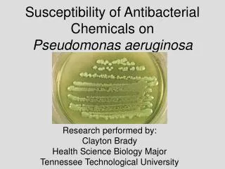
Other infections associated with contaminated water or liquid solutions • Outer ear infections (swimmers ear) • Eye infections (contact lens wearers especially) • Endocarditis (IV drug abusers) • Treatment Challenges • Multiple antibiotic resistance problems • Develops resistance overnight
Mechanisms of Antibiotic Resistance in Pseudomonas aeruginosa
LAB DIAGNOSIS • Specimen – Pus, wound swab, urine, catheter tip, ET aspirate • Microscopy – Long slender GNB, Motile • Culture – NA – greenish pigmentation • BA – greyish haemolytic • MA – NLF • Cetrimide agar – selective media
Biochemical reactions • Oxidase & Catalase +ve • Non fermenter • Oxidative breakdown of Glucose • Nitrate Nitrite Nitrogen • Antibiotic senstivity – Piperacilin, carbencillin, Amikacin, Meropenm • Pyocin typing – R, F & S • 105 types • Serotyping – O & H Antigens; 17 serotypes
Burkholderia • They colonize the moist environmental surfaces and are commonly associated with nosocomial infections. • B. cepacia complex (of 9 species), B. gladioli and B. pseudomallei are important pathogens. • B. cepacia complex causes RT infections particularly in cystic fibrosis patients, UT infections and septicemia. • Usually non-fatal except for RT infections in CF patients.
B. pseudomallei usually causes opportunistic infections (called melioidosis), but may sometimes infect previously healthy persons. • Infection by this organism may result in asymptomatic infection, acute suppurativecutaneous infection, and chronic pulmonary infection ranging in severity from mild bronchitis to necrotizing pneumonia. All may progress to sepsis.
Stenotrophomonasmaltophilia • A common nonfermentative, gram-negative isolate. • It infects debilitated or immunocompromised persons, and causes a wide spectrum of diseases, including wound infections, UT infections, pneumonia, sepsis, meningitis, etc. • It is resistant to many commonly used antibiotics, and patients receiving long-term antibiotic therapy are particularly at risk for acquiring infections. • Infections may be acquired from iv catheters, contaminated disinfectants, respiratory therapy and monitoring equipment, and ice machines.
Surgical incision is given Wound is cleaned
Surgical Care: • Opening the wound • Evacuating pus • Cleansing the wound • The deeper tissues are inspected for a deep space infection • Frequent dressing change
How to classify? • 1st Degree (Epidermis): low risk of infection • 2nd Degree (Dermis): Low – moderate risk • 3rd Degree (Dermis): High risk
Zone of necrosis Edema Zone of injury Normal tissue • Characteristics • Necrosis to upper third of dermis • Zone of necrosis lifted off viable wound by edema • 3. Small zone of injury
Characteristics • Necrosis to majority of skin • Zone of necrosis adherent to zone of injury • 3. Smaller edema layer
Characteristics • No remaining viable dermis
Microbiology • Organisms most frequently isolated from burn wound biopsy specimens • Staphylococcus aureus • Pseudomonas aeruginosa • Enterobacter cloacae • Klebsiella pneumoniae • Enterococcus faecalis • Acinetobacter baumannii • Aspergillus species • Candida albicans
Lab diagnosis • Staining methods • Gram stain for infective organisms • Staining for fungal elements • Culture techniques • Both aerobic and anaerobic organisms • Fungal cultures • Drug sensitivity testing • Newer techniques • ELISA or RIA for detection of Ag/Ab • Detection of RNA or DNA sequences or protein by Northern, Southern or Western blotting, respectively • PCR