Download
1 / 25
821 likes | 1.91k Views
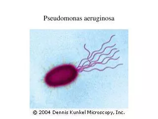
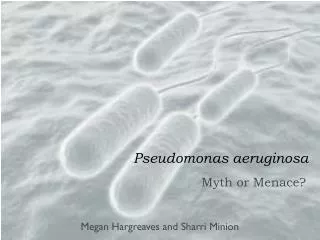
PSEUDOMONAS AERUGINOSA. Pseudomonas aeruginosa. Gram-negative bacillus, polar flagella Pigmented growth on agar and distinct grape like odor usually green or brown, smells like grapes or juicy fruit gum Ubiquitous in environment, especially in water

E N D
Pseudomonas aeruginosa • Gram-negative bacillus, polar flagella • Pigmented growth on agar and distinct grape like odor • usually green or brown, smells like grapes or juicy fruit gum • Ubiquitous in environment, especially in water • Hospital environment loaded with this pathogen • Hardy, survive extreme conditions • PRIMARILY OPPORTUNISTIC PATHOGEN
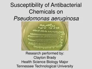
Pigmented P.aeruginosa P.aeruginosa on blood agar
Pigment production: • Pyocyanin: bluish green, soluble in water & chloroform • Fluorescein: (pyoverdin): greenish yellow- soluble only in chloroform • Pyorubin • Pyomelanin • BLUE PUS
Pseudomonas aeruginosa • Virulence Factors • Attachment by fimbriae: Pili and nonpilus adhesins • Capsule/ slime is anti-phagocytic: Capsule (alginate, glycocalyx): seen in cultures from patients with cystic fibrosis. • Endotoxin – inflammation: LPS
P.aeruginosa - Virulence Factors • Exotoxin A • Blocks protein synthesis • Kills cells • Skin or lung damage depends on infection • Pyocyanin: catalyzes production of toxic forms of oxygen that cause tissue damage. It also induces IL-8 production. • Pyoverdin: a siderophore.
Proteases: Serine protease, metalloprotease, alkaline protease cause tissue damage and help bacteria spread. • Phospholipase C: a hemolysin • Exotoxin A: causes tissue necrosis and is lethal for animals (disrupts protein synthesis); immunosuppressive. • Exoenzyme S and T: cytotoxic to host cells.
Infections caused by P.aeruginosa • COMMUNITY ACQUIRED INFECTIONS • Suppurative otitis media • Respiratory tract infections in cystic fibrosis pts. NOSOCOMIAL INFECTIONS: • Urinary tract infections • Infection of burn wounds, bedsores, surgical site • Septicemia, endocarditid, meningitis, • Ventilator assoc. pneumonia
Opportunistic infections • Immunocpmromised host • Colonize hospitalized patients via • Respirators • IV solutions • Instruments • Cystic fibrosis patients • Burn victims are at particular risk to rapidly lethal infections.
Pulmonary Infections • Colonization • Benign bronchitis to necrotic fatal pneumonia • Cystic Fibrosis • Mucoid encapsulated strains are major problem • Exacerbations of disease happen frequently • Invasive pneumonia • Neutropenic patients • Respiratory equipment is a primary source • Serious pneumonia
Skin Infections • Most common in burn victims • Moist environment • Lack of WBC reaching damaged tissue • Vascular damage • Tissue necrosis • Blood stream invasion • Folliculitis (hot tubs, whirlpools, pools)
Infections associated with contaminated water or liquid solutions • Outer ear infections (swimmers ear) • Eye infections (contact lens wearers especially) • Endocarditis (IV drug abusers) • Treatment Challenges • Multiple antibiotic resistance problems • Develops resistance overnight
Mechanisms of Antibiotic Resistance in Pseudomonas aeruginosa
LAB DIAGNOSIS • Specimen – Pus, wound swab, urine, catheter tip, ET aspirate • Microscopy – Long slender GNB, Motile • Culture – Nutrient agar – greenish pigmentation • Blood agar: greyish haemolytic • Mac conkey agar: Non Lactose Fermenting • Cetrimide agar – selective media
Biochemical reactions • Oxidase & Catalase +ve • Non fermenter • Oxidative breakdown of Glucose • Nitrate Reduction test: Positive • Antibiotic senstivity – Piperacilin, carbencillin, Amikacin, Meropenm • Pyocin typing – R, F & S • 105 types • Serotyping – O & H Antigens; 17 serotypes
Burkholderia • Colonize moist environmental surfaces and are commonly associated with nosocomial infections • B.cepacia; oppurtunisticenvironmatal pathogen • In patients with cystic fibrosis & chronic granulomatous diseases • Fatal necrotizing pneumonia • Nutritionally very versatile • Causes: urinary, respiratory, wound infections • Inherently resistant to most antibiotics
B. pseudomallei: • Causes opportunistic infection (called melioidosis), but may sometimes infect previously healthy persons • Glanders like disease • First described by Whitmore & Krishnaswami • Infection by this organism may result in asymptomatic infection, acute suppurativecutaneous infection, and chronic pulmonary infection ranging in severity from mild bronchitis to necrotizing pneumonia. All may progress to sepsis.
Infection acquired by: contamination of wounds with soil and water • Common in diabetics • It may take years for the disease manifests. Long latency: REACTIVATION • Acute infection: Acute septicemia, subacute typhoid like disease, pneumonia. High fatality rate • Chronic infection: Multiple caseous, suppurative foci, abscess formation in s/c tissue, bones andinternalorgans
DIAGNOSIS: • Microscopy: demonstration of small irregular staining GNB showing bipolar safety pin appearance: when stained with methylene blue stain • Serology: ELISA for IgM and IgG • PCR test
Treatment • Ceftazidime: drug of choice • Cotrimoxazole, tetracycline, amoxycillin, chloramphenicol • Prolonged treatment
Stenotrophomonasmaltophilia • A common non-fermentative, gram-negative bacteria • It infects debilitated or immunocompromised persons, and causes a wide spectrum of diseases, including wound infections, UT infections, pneumonia, sepsis, meningitis, • Resistant to many commonly used antibiotics, and patients receiving long-term antibiotic therapy are particularly at risk for acquiring infections. • Infections may be acquired from IV catheters, contaminated disinfectants, respiratory therapy and monitoring equipment, and ice machines.
Glanders • B.mallei • Non motile gram negative bacilli • Causes GLANDERS : a disease of horses & mules • Human infection: occupational • Acute/ chronic infection of respiratory tract, skin subcutaneous tissue • Laboratory cultures are highly infectious