Download
1 / 63
630 likes | 842 Views
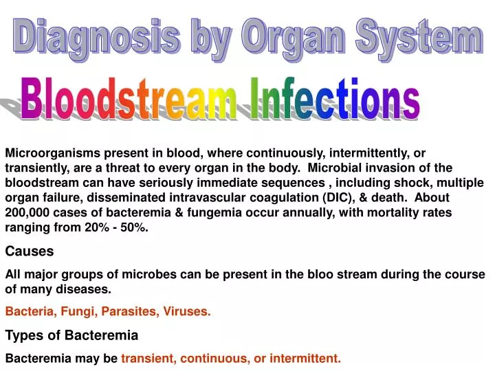
Diagnosis by Organ System. Bloodstream Infections.

E N D
Diagnosis by Organ System Bloodstream Infections Microorganisms present in blood, where continuously, intermittently, or transiently, are a threat to every organ in the body. Microbial invasion of the bloodstream can have seriously immediate sequences , including shock, multiple organ failure, disseminated intravascular coagulation (DIC), & death. About 200,000 cases of bacteremia & fungemia occur annually, with mortality rates ranging from 20% - 50%. Causes All major groups of microbes can be present in the bloo stream during the course of many diseases. Bacteria, Fungi, Parasites, Viruses. Types of Bacteremia Bacteremia may be transient, continuous, or intermittent.
Types of Bloodstream Infections 1- Intravascular: Those that originate within the cardiovascular system. A- Endocarditis. B- Mycotic aneurysm C- Suppurative thrompophlepitis. D- Intravenous catheter-associated bacteremia. 2- Extravascular:Those that result from bacteria entering the blood circulation through the lymphatic system from another site of infection. Most cases are a result of extravascular infection. The common portals of entry for bacteremia are: most 1- The genitourinary tract (25%). 2- Respiratory tract (20%). 3- Abscesses (10%). 4- Surgical wound infections (5%). 5- Biliary tract (5%). 6- Miscellaneous sites (10%), & uncertain sites (25%).
Clinical Manifestations Bacteremia: Indicates the presence of a focus of disease, such as intravascular infection, pneumonia, or liver abscess, or transient release of bacteria into bloodstream. Septicemia or sepsis: Indicates a situation in which bacteria or their products (toxins) are causing harm to the host. Unfortunately clinicians often use these terms interchangeably. Symptoms may include: fever or hypothermia, chills, hyperventilation (abnormal increased breathing that leads to excess loss of CO2 from the body) & subsequent alkalosis (condition caused by the loss of acid leading to an increase in pH), skin lesions, change in mental status, & diarrhea. More serious manifestations include hypotension or shock, DIC & major organ failure. The syndrome known as septic shock, characterized by fever, acute respiratory distress, shock, renal failure, intravascular coagulation, & tissue destruction, can be initiated by either endotoxins or exotoxins. Septic shock is mediated by activated mononuclear cells producing cytokines, such as tumor necrosis factor & interleukins.
Immunocompromised Patients Detection of Bacteremia Mortility rate associated with bacteremia ranges from 20% - 50 %. 1- Specimen Collection A- Preparation of the site. B- Antisepsis. C- Precautions. D- Specimen volume Adults: 10 -20 ml is recommended. Children: 1 – 5 ml. Infants & small E- Number of blood cultures: At least 3 blood cultures to rule out negative results. F- Timing of collection: When there is symptoms, any time. G- Anticoagulation & dilution. H- Blood culture media & additives.
2- Culture Techniques A- Conventional blood cultures 1- Incubation conditions. 2- Detecting growth. 3- Self-contained subculture system. 3- Lysis centrifugation. 4- Instrument – based systems – Bactec systems, BacT/ALERT microbial detection system, ESP system. 5- Techniques to detect IV catheter – associated infections. 6- Handling positive blood cultures. 7- Interpretation of blood culture results. Special considerations for other relevant organisms isolated from blood Hacek bacteria Fungi Mycobacteria Spirochetes, Mycoplasma hominis, Bartonella,B6 dependent Streptococci.
Anatomy Diseases of the Lower Respiratory Tract 1- Bronchitis. A- Acute: usually caused by viruses. In infants & preschool children is Bordetella pertussis. B- Chronic: caused by bacteria, such noncapsulated strains of Haemophilus influenzae, Streptococcus pneumoniae & Moraxella catarrhalis. 2- Pneumonia Pathogenesis Clinical Manifestations Community-Acquired Pneumonia: patients are believed to have acquired infection outside the hospital setting. The etiology of acute pneumonias is strongly dependent on age. More than 80% of pneumonias in infants & children are caused by viruses, whereas 10%-20% of pneumonias in adults are viral.
Children Children suffer less commonly from bacterial pneumonia, usually caused by H. influenzae, S. pneumoniae, or S. aureus. Neonates may acquire lower respiratory tract infections with C. trachomatis or P. carinii. Young Adults The most common etiologic agent of pneumonia in adults <30 years age is Mycoplasma pneumoniae which is transmitted via close contact. Adults Community acquired pneumonia in adults is most commonly due to bacterial infections. S. pneumoniae is most prevalent, causing 80% of all community-acquired pneumonia. Pneumonia secondary to aspiration of gasric or oral secretions is common &occurs in the community setting. Hospital-Acquired Pneumonia It is the leading cause of death among patients with nosocomial infections (as high as 50% mortality among patients in intensive care units). The most common etiologic agents include Klebsiella spp., other Enterobacteriaceae, S. aureus, anaerobes, S. pneumoniae, P. aeruginosa, & Legionella. Viruses as influenza virus, respiratory syncytial virus, & adenovirus.
Chronic Lower Respiratory Tract Infections Mycobacterium tuberculosis is the most likely etiologic agent of chronic lower respiratory tract infection, but fungal infection & anaerobic pleuropulmonary infection may also run a subacute or chronic course. Cystic fibrosis (CF) is a genetic disorder that leads to persistent bacterial infection in the lung, causing airway wall damage & chronic obstructive lung disease. Immunocompromised Patients Patients with neoplasm Transplant recipients HIV-infected patients Pleural Infections Laboratory Diagnosis of Lower Respiratory Tract Infections Specimen Collection & Transport Sputum, transtracheal aspirate, bronchial washings. Specimen Processing Direct visual Examination Routine Culture
Upper Respiratory Tract Infections & Other Infections of the Oral Cavity
Laryngitis Caused almost exclusively by viruses. Laryngotracheobronchitis or Croup Virusesa primary cause of croup. Epiglottitis Is usually caused by bacteria. Haemophilus influenzae type b is the primary cause of epiglottitis. Other organisms occasionally implicated are streptococci & staphylococci. Pharyngitis & tonsillitis Pharyngitis (sore throat) & tonsillitis are common upper respiratory tract infections affecting both children & adults. Clinical Manifestations Affected tissues are erythematous & swollen with pain. Depending on the causative organism, either inflammatory exudates (fluid with protein, inflammatory cells, & cellular debris), vesicles (small blister like sacs containing liquid) & mucosal ulceration, or nasopharyngeal lymphoid hyperplasia (swollen lymph nodes) may be observed.
Etiologic agents Most cases of pharyngitis occur during the colder months & often accompany other infections, primarily those caused by viruses. Although different bacteria cause pharyngitis & tonsillitis, the primary cause is Streptococcus pyogenes. Although H. influenzae, S. aureus, & S. pneumoniae are frequently isolated from nasopharyngeal & throat cultures, they have not been shown to cause pharyngitis. Vincent's angina or anaerobic tonsillitis, involves pseudomembrane formation on tonsillar surfaces. Multiple anaerobes, especially Fusobacterium necrophorum are implicated in this syndrome. Gram stain reveals numerous fusiform, gram-negative bacilli & spirochetes. Peritonsillar Abscesses Caused by non-spore-forming anaerobes, includingFusobacterium, Bacteroides, &anaerobic cocci. S. pyogenes & viridans streptococci may also be involved. Rhinitis (common cold) Inflammation of nasal mucous membrane or lining. It is caused by viruses.
Miscellaneous Infections Caused by Other Agents Corynebacterium diphtheriae Bordetella pertussis Klebsiella spp. Rhinoscleroma is a rare form of chronic, granulomotous infection of the nasal passages, including the sinuses & occasionally the pharynx & larynx. Associated withK. rhinoscleromatous, the disease is associated with nasal obstruction caused by tumorlike growth with local extension. K. Ozaenae may contribute to another infrequent condition called ozena, characterized by a chronic, mucopurulent nasal discharge that is often foul-smelling. It is caused by secondary, low-gradeanaerobic infection. Oral Cavity Stomatitis Herpes simplex virus agent of the disease, in which multiple ulcerative lesions are seen on the oral mucosa. is the primary. Thrush Caused by Candida spp.
Periodontal Infections It involves anaerobes, Streptococci, Staphylococci & Eikenella corrodens. Salivary Glands Infections Staphylococcus aureusis the major pathogen, but viridans streptococci & anaerobes may play a role. Mumps virus Neck Diagnosis of the Upper Respiratory Tract Infections Collection & Transport of Specimens Dacron, cotton, or calcium alginate-tipped swabs are suitable for collecting most upper respiratory tract microorganisms. Moist swabs can remain for 4 hours, otherwise transport medium is needed S. pyogenes is an exception as it remains for 48-72 hours viable in dry swabs. Nasopharyngeal swabs are suitable for detecting other bacteria & viruses (Bordetella pertussis, Neisseria spp.) Direct Visual Examination Gram stain is of little help. 10% KOH for fungi
Culture Streptococcus pyogenes Corynebacterium diphtheriae Bordetella pertussis Neisseria Epiglottitis Noncutlure Methods for Detection of S. pyogenes in Throat Specimens. Diagnosis of Infections in the Oral Cavity & Neck Collection & Transport Direct Visual Examination Culture
Meningitis& Other Infections of the CNS Anatomy An understanding of the basic anatomy & physiology of the CNS is helpful for the microbiologist to ensure appropriate specimen processing & interpretation of laboratory results. Coverings & spaces of the CNS 1- Bone outercovering of the brain & the spinal cord. 2- Inner covering of membranes called meninges which are of 3 layers surrounding the brain & the spinal cord. These are: A- Dura mater B- Arachnoid C- Pia mater The last 2 are collectively called leptomeninges. Between & around the meninges are spaces that include the epidural, subdural, & subarachnoid spaces.
Cerebrospinal Fluid Routes of Infection Diseases of the CNS Meningitis Infection within the subarachnoid space or throughout the leptomeninges is called meningitis. It is divided into 2 major categories 1- Purulent Meningitis Bacteria usually cause these infections. Clinical Manifestations Acute Chronic Etiologic Agents It is very dependent on the age of the patient. H. Influenzae is the most etiologic agent in children between 1 month & 6 years. About 95% of the cases are caused by Hib, S. pneumoniae, & N. meningitidis.
Neonates are likely to be infected by S. agalactiae, E. coli, & other gramnegative bacilli & Listeria monocytogenes. Occasionally other organisms are involved, Chrysobacterium meningosepticum. In adults N. meningitidis, S. pneumoniae,, S. aureus & various gram negative bacilli. 2- Aseptic Meningitis Commonly associated with viral infection. Encephalitis/Meningoencephalitis An inflammation of the brain parenchyma & is usually a result of viral infection. Brain Abscess Laboratory Diagnosis Meningitis Specimen collection & Transport CSF is collected aseptically by inserting a needle into the subarachnoid space at the level of the lumbar spine. A minimum of 5-10 ml should be collected. .
Specimen should be delivered immediately to the laboratory. Specimen should not be refrigerated. If specimen is not processed immediately, it should be incubated at 35c or left at room temperature. Initial Processing Microbiology 1- Direct stained smear 2- Wet preparation 3- India ink stain 4- Direct Detection of etiologic agents. 5- Culture Cytology Total cell count & differential. Biochemistry Glucose & protein level.
Infections of the Eye, Ear & Sinuses Eye Anatomy Resident Microbial Flora Staphylococcus epidermidis, & Lactobacillus spp. are the most frequently encountered organisms, Propionibacterium acnes, S. aureus, H. influenzae, Moraxella catarrhalis, Enterobacteriaceae & various Streptococci. Diseases Blephritis Conjuncivitis Keratitis Endophthelmitis Periocular
Laboratory diagnosis Specimen Collection & Transport Purulent material from the surface of the lower conjunctival sac & inner canthus (angle) of the eye is collected on a sterile swab for culture of conjunctivitis. Both eyes should be cultured separately. In keratitis, an ophthalmologist should obtain scrapings of the cornea with a heat sterilized platinum spatula. Cultures of endophthalmitis specimens are inoculated with material obtained by the ophthalmologist from the anterior & posterior chambers of the eye. Lid infection material is collected on a swab Direct Visual Examination Gram stain, DFA Culture Blood agar, chocolate agar. Incubation under 5-10% CO2 at 37c for 24-48 hors. Noneculture method ELISA, DFA, & PCR for Chlamydia trachomatis & viruses.
Ears Anatomy Resident Microbial Flora The normal flora of the external canal are rather sparse, similar to that of the conjunctival sac qualitatively. Staphylococcus epidermidis, & Lactobacillus spp. are the most frequently encountered organisms, Propionibacterium acnes, S. aureus, H. influenzae, Moraxella catarrhalis, Enterobacteriaceae & various Streptococci. Diseaes Otitis Externa Otitis Media Laboratory Diagnosis Specimen Collection & Transport
Ear discharge. Culture Media: blood agar, chocolate agar, MacConkey. Sinuses Anatomy Diseases Laboratory Diagnosis Gram stain Aerobic Culture: blood agar, chocolate agar, MacConkey. Anaerobic Culture
Infections of the urinary Tract Anatomy
Resident Flora The urethra has resident microflora the colonize its epithelium in the distal portion. All areas of the urinary system above the urethra in a healthy human are sterile. Coagulase negative staphylococci, diphtheroids, viridans & nonhemolytic streptococci, lactobacilli, nonpathogenic Neisseria, anaerobic gram negative cocci & bacilli, & commensal Mycobacteria & Mycoplasma spp. Infection of the Urinary Tract Epidemiology Approximately 10% of humans will have a UTI at some time during their lives. It is age & sex dependent. During the first year of life UTIs are more common in males. However, the incidence of UTIs among males is low after age 1& until approximately age 60 when enlargement of the prostate interferes with emptying of the urinary bladder. Therefore UTI is predominantly a disease of females. Sexual activity, anatomic & hormonal changes 7 pregnancy favor development of UTIs. Etiologic Agents 1- Community Acquired 2- Hospital Acquired
Types of Infections & their Manifestations 1- Urethritis 2- Cystitis 3- Acute Urethral Syndrome 4- Pyelonephritis Laboratory Diagnosis of Urinary Tract Infections Specimen Collection Clean-Catch, Midstream Urine Straight Catheterized Urine Suprapubic Bladder Aspiration Indwelling Catheter Specimen Transport Screening Procedures Gram stain, pyuria, indirect indices, nitrate reductase, leukocytes esterase, catalase. Automated systems.
Urine Culture Inoculation & Incubation of Urine Cultures Interpretation of Urine Cultures
Gastrointestinal Tract Infections Anatomy Resident Microbial Flora Gastroenteritis Pathogenesis Host Factors Microbial factors Toxins Entertoxins Cytotoxins Neurotoxins Clinical Manifestations
Other Infections of the GI Tract Esophagitis The most common etiologic agents are Candida spp., herpes simplex & CMV. Gastritis Helicobacter pylori. Procitis Chlamydia trachomatis, herpes simplex, syphilis, & gonorrhea are the most common etiologic agents. Miscellaneous Laboratory Diagnosis of Gastrointestinal Tract Infections Specimen Collection & Transport Stool specimens should be collected in clean plastic container & delivered to the laboratory within one hour. Stool for toxin assay for Clostridium difficile, immunoelectron microscopy for rotavirus, ELISA or latex agglutination for rotavirus should be sent without preservatives.
Stool Specimen for Bacterial Culture If a delay more than 2 hours is anticipated for stools for culture, the specimen should be placed in transport medium such as Cary-Blair transport medium. If stool is not available, a rectal swab may be substituted as a specimen for bacterial or viral culture. Stool Specimens for Viruses Miscellaneous Specimen Types Direct Detection of Agents of Gastrointestinal Types Wet Mounts Stains Antigen Detection Molecular Biological Techniques Culture of Fecal Material for Isolation of Etiologic Agents Routine Culture
A C E
Skin, Soft Tissue, & Wound Infections Anatomy of Skin Etiologic Agents & Pathogenesis Many different bacteria, fungi 7 viruses may be involved. Skin & Soft Tissue Infection Skin Infections in or around Hair Follicles Folliculitis, furnculosis, & carbuncles are localized abscesses either in or around hair follicles. Staphylococcus aureus is the most etiologic agent for all three infections. Infections in the Keratinized Layers of the Epidermis Because of their ability to utilize keratin in the cells of the epidermis, hair & nails, the dermatophyte fungi are significant & well suited pathogens for this site. Infections of the Deeper Layers of the epidermis &the Dermis
Infections of the Subcutaneous Tissues Abscesses, ulcers & boils. S. aureus is the most common etiologic agent of subcutaneous abscesses in healthy individuals. Also the etiologic agent depends on the site of infection. Infections of the Muscle Fascia & Muscles Necrotizing Fascitis Progressive Bacterial Synergistic Gangrene Myositis Wound Infections Postoperative Infections Bites Burns Infections Related to Vascular & Neurologic Problems Sinus Tract & Fistulas Systemic Infections with Skin Manifestations
Laboratory Diagnostic Procedures Infections of the Epidermis & Dermis Erysipeloid Superficial Mycosis & Erythrasma Erysipelas & Cellulitis Vesicles 7 Bullae Infections of the Subcutaneous Tissues Infections of the Muscle Fascia & Muscles Wound Infections Postoperative Bites Burns