Download
1 / 50
520 likes | 571 Views
Infectious Arthritis. N.Movaffagh MD Rheumatologist. Acute bacterial infection typically involves a single joint or a few joints. chronic monarthritis or oligoarthritis suggests: mycobacterial or fungal infection episodic inflammation suggests:

E N D
Infectious Arthritis N.Movaffagh MD Rheumatologist
Acute bacterial infection typically involves a single joint or a few joints
chronic monarthritis or oligoarthritis suggests: • mycobacterial or fungal infection • episodic inflammation suggests: • syphilis, reactive arthritis
Acute polyarticular inflammation: • Endocarditis rheumatic fever disseminated neisserial infection acute hepatitis B • Bacteria and viruses occasionally infect multiple joints ( eg …rheumatoid arthritis)
PATHOGENESIS • Bacteria enter the joint from: • Bloodstream • contiguous site of infection in bone or soft tissue • direct inoculation during surgery • injection, animal or human bite • trauma
In hematogenous infection: • bacteria escape from synovial capillaries,(have no limiting basement membrane) • provoke neutrophilic infiltration of the synovium. • Neutrophils and bacteria enter the joint space bacteria adhere to articular cartilage • Degradation of cartilage begins with in 48 h • result of increased intraarticular pressure release of proteases and cytokines from chondrocytes and synovial macrophages
bacteria lining the synovium and cartilage abscesses extending into the synovium, cartilage, and in severe cases subchondral bone • Thrombosis of inflamed synovial vessels
Bacterial factors in the pathogenesis: • surface-associated adhesinsin S. aureus • Endotoxins(promote chondrocyte-mediated breakdown of cartilage)
MICROBIOLOGY • hematogenous route of infection is the most common route in all age
most common pathogens • In infants group B streptococci, gram-negative enteric bacilli, S. aureus • children <5 years of age • S. aureus, Streptococcus pyogenes (group A Streptococcus) and (in some centers) Kingellakingae
young adults and adolescents: • N. gonorrhoeae • adults of all ages: • S. aureus accounts for most nongonococcalisolates • older adults • gram-negative bacilli, pneumococci, β-hemolytic streptococci groups A and B β-hemolytic streptococci C, G, F , in especially those with underlying comorbid illnesses
Infections after surgical procedures or penetrating injuries are suggestive: • S. aureusand occasionally to other gram-positive bacteria or gram-negative bacilli • after the implantation of prosthetic joints or arthroscopy are suggestive: • coagulase-negative staphylococci
after human bites and when decubitus ulcers or intraabdominal abscesses: • Anaerobic organisms often in association with aerobic bacteria • AfterBites and scratches from cats and other animals: • Pasteurella multocida or Bartonella henselae
Penetration of a sharp object through a shoe: • Pseudomonas aeruginosa arthritis in the foot
NONGONOCOCCAL BACTERIAL ARTHRITIS • Epidemiology: • RA have the highest incidence of infective arthritis (most often secondary to S. aureus) • because of chronically inflamed joints; glucocorticoid therapy; frequent breakdown of rheumatoid nodules, vasculitic ulcers, and skin overlying deformed joints.
Diabetes mellitus, glucocorticoid therapy, hemodialysis, malignancy carry an increased risk of infection with S. aureus and gram-negative bacilli • Tumor necrosis factor inhibitors predispose to mycobacterial infections and other pyogenic bacterial infections
alcoholism, deficiencies of humoral immunity, and hemoglobinopathies associated Pneumococcal infections • Pneumococci, Salmonella species, H. influenzae • cause septic arthritis in HIV • primary immunoglobulin deficiency are at risk for mycoplasmal arthritis
IV drug users acquire: staphylococcal and streptococcal infections from their own flora pseudomonal and other gram-negative infections from drugs and injection paraphernalia
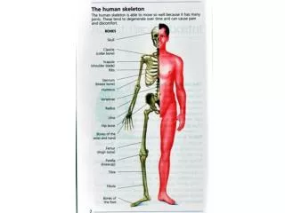
Clinical Manifestations • Some 90% of patients present with Involvement of a single joint most commonly the knee hip; and still less often the shoulder, wrist, elbow • Small joints of the hands and feet are more be affected after direct inoculation or a bite • infections of the spine, sacroiliac joints, and sternoclavicular joints are more common in IV drug users
Polyarticular infection is most common in rheumatoid arthritis and may resemblea flare of the underlying disease
Clinical Manifestations • pain • Effusion • muscle spasm • decreased ROM • Fever ( 38.3–38.9°C) • {may not be present, especially in RA,renalor hepatic insufficiency, or conditions requiring immunosuppressive therapy}
DD • Cellulitis • Bursitis • acute osteomyelitis • extraarticular infection, such as a boil or pneumonia,should be sought
Imaging • Plain radiographs: • soft-tissue swelling • Joint space widening • displacement of tissue planes by the distended capsule • advanced infection: • Narrowing of the joint space • bony erosions
Imaging • Ultrasound is useful for detecting effusions in the hip • CT or MRI can demonstrate infections of the sacroiliac joint, sternoclavicular joint, spine
Laboratory Findings • leukocytosis with a left shift • elevation of the ESR or CRP • Blood cultures are positive in up to 50–70% of S.aureus infections
Synovial fluid • Turbid • serosanguineous • Frankly purulent • large numbers of neutrophils (in Gram-stained smears) • Elevation of total protein and LDH not specific • glucose level not specific
Positive smears in synovial fluid: three-quarters of infections with S. aureus and streptococci • 30–50%of infections due to gram-negative and other bacteria
Cultures of synovial fluid are positive in >90% • NAA-based assays for bacterial DNA, can be useful for the diagnosis of partially treated or culture-negative bacterial arthritis.
Normal synovial fluid contains <180 cells (predominantly mononuclear cells) per microliter • Synovial cell counts averaging 100,000/μL (range, 25,000–250,000/μL), with >90% neutrophils, are characteristic of acute bacterial infections
cell counts <30,000–50,000 cells/μL: • Crystal-induced, rheumatoid, and other noninfectious inflammatory arthritides • cell counts 10,000–30,000/μL, with 50–70% neutrophilsand the remainder lymphocytes: • mycobacterial and fungal infections
Definitive diagnosis of an infectious process: • 1.identification of the pathogen in stained smears of synovial fluid • 2.isolation of the pathogen from cultures of synovial fluid and blood • 3.detection of microbial nucleic acids and proteins by nucleic acid amplification (NAA)–based assays and immunologic techniques
TREATMENT Bacterial Arthritis • Empirical antibiotics against the bacteria visualized on smears or pathogens based on age and risk factors • Drainage of the involved joint • Arthroscopic drainage and lavage • Arthrotomy
IV third-generation cephalosporin such as cefotaxime (1 g every 8 h) or ceftriaxone (1–2 g every 24 h) • IV vancomycin(1 g every 12 h) is used if there are gram-positive cocci on the smear • oxacillin or nafcillin(2 g every 4 h) • If methicillin-resistant S. aureus is an unlikely pathogen
aminoglycoside or third generationcephalosporin should be given to IV drug users (P. aeruginosa)
Infections due to staphylococci are treated with oxacillin, nafcillin, or vancomycin for 4 weeks.
do not require immobilization except for pain control before symptoms are alleviated by treatment • Weight bearing should be avoided until signs of inflammation have subsided • Frequent passive motion of the joint is indicated
GONOCOCCAL ARTHRITIS • incidence has declined in recent years • consequence of bacteremia arising from gonococcal infection or, more frequently,from asymptomatic gonococcal mucosal colonization of the urethra,cervix, or pharynx • Women are at greatest risk during menses and during pregnancy • two to three times more likely than men to develop disseminated gonococcal infection (DGI) and arthritis
up to 70% of episodes of infectious arthritis in persons <40 years of age in the United States
GONOCOCCAL ARTHRITIS 1.Disseminated gonococcal infection (DGI) 2.Gonococcal septic arthritis
DGI • most common manifestation of DGI: • fever, chills, rash, and articular symptoms • Papules ,hemorrhagic pustules the trunk and the extensor surfaces of the distal extremities • Migratory arthritis and tenosynovitisof the knees, hands,wrists, feet, and ankles are prominent
Causes of cutaneous lesions and articular findings: • reaction to circulating gonococci and immune-complex deposition in tissues • cultures of synovial fluid are consistently negative • blood cultures are positive in fewer than 45% • Synovial fluid : 10,000–20,000 leukocytes/μL
Gonococcalseptic arthritis • less common than the DGI syndrome • always follows DGI • A single joint such as the hip, knee, ankle, or wrist • Synovial fluid, contains >50,000 leukocytes • G is evident only occasionally in Gram-stained smears, • cultures of synovial fluid are positive in fewer than 40% • Blood cultures are almost always negative
NAA-based urine tests also may be positive • NAA-based assays are extremely sensitive(in synovial fluid) • Cultures and Gram-stained smears of skin lesions Occasionally positive. • All specimens for culture should be plated onto Thayer-Martin agar • Or special transport media at the bedside and transferred promptly to the microbiology laboratory in an atmosphere of 5% CO2,
A dramatic alleviation of symptoms within 12–24 h after the initiation of appropriate antibiotic therapy • treatment consists of ceftriaxone • Then 7-day course of therapy an oral fluoroquinolone • Patients with DGI should be treated for Chlamydia trachomatis infectionunless this infection is ruled out by appropriate testing
INFECTIONS IN PROSTHETIC JOINTS • Majority of infections are acquired intraoperatively or immediately postoperatively • Presentation may be acute: • fever, pain, and local signs of inflammation, especially due to S. aureus, pyogenic streptococci, and enteric bacilli
coagulase-negative staphylococci or diphtheroids: persist for months or years without causing constitutional symptoms • They acquired during joint implantation or discovered during evaluation of chronic unexplained pain or after a radiograph shows loosening of the prosthesis C-reactive protein ESR
Diagnosis: • needle aspiration of the joint; Sonication of explanted prosthetic material can improve the yield of culture • by breaking up bacterial biofilms on the surfaces of prostheses • if routine and anaerobic cultures are negative: • Use of special media for such as fungi, atypical mycobacteria, and Mycoplasma
Treatment • high doses of parenteralantibiotics&surgery(4–6 weeks) • In most cases, the prosthesis must be replaced • In some cases, reimplantation is not possible, and the patient must manage without a joint
Cure of infection without removal of the prosthesis: • in streptococci or pneumococciand that lack radiologic evidence of loosening of the prosthesis • Antibiotic therapy&joint should be drained by open arthrotomy or arthroscopically • oral rifampin and ciprofloxacin for 3–6 months to persons with staphylococcal prosthetic joint infection