Download
1 / 32
320 likes | 456 Views
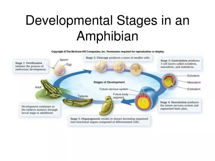
Developmental Stages in an Amphibian. Animal development. Gut. Cell movement. Zygote (fertilized egg). Eight cells. Blastula (cross section). Gastrula (cross section). Adult animal (sea star). LE 21-4. Cell division. Morphogenesis. Observable cell differentiation. Seed leaves.

E N D
Animal development Gut Cell movement Zygote (fertilized egg) Eight cells Blastula (cross section) Gastrula (cross section) Adult animal (sea star) LE 21-4 Cell division Morphogenesis Observable cell differentiation Seed leaves Plant development Shoot apical meristem Root apical meristem Two cells Zygote (fertilized egg) Embryo inside seed Plant
Most animals proceed • through these stages • during embryonic • development: • Zygote • Early cleavage stages • Morula (solid ball) • Blastula (hollow ball) • Gastrula
LE 47-7 Morula Fertilized egg Blastula Four-cell stage
Starfish development, unfertilized egg. 4 blastomeres. 2 blastomeres. Starfish development, nonmotile blastula. 16 blastomeres. 32 blastomeres. morula
Unfertilized egg cell Sperm Molecules of another cytoplasmic determinant Molecules of a cytoplasmic determinant Nucleus • Egg provides proteins and mRNAs required for early development • Cleavage asymmetrically divides cytoplasmic components; immediately establishing polarity Fertilization LE 21-11a Zygote (fertilized egg) Mitotic cell division Two-celled embryo Cytoplasmic determinants in the egg
Animal hemisphere Animal pole Point of sperm entry Vegetal hemisphere LE 47-8 Vegetal pole Point of sperm entry Future dorsal side of tadpole Anterior Gray crescent Right First cleavage Ventral Dorsal Left Posterior Body axes Establishing the axes
Zygote 0.25 mm 2-cell stage forming LE 47-9 4-cell stage forming Eight-cell stage (viewed from the animal pole) 8-cell stage 0.25 mm Animal pole Blasto- coel Blastula (cross section) Vegetal pole Blastula (at least 128 cells)
Gastrulation - Establishing Germ Layers (tissue development) • Ectoderm gives rise to outer covering and nervous system • Endoderm gives rise to the digestive tract • Mesoderm gives rise to muscle tissue
Starfish development, gastrula during invagination. Starfish development, mid-gastrula. LM X75. Starfish, late bipinnaria. Starfish, young adult.
Neurulation • The nervous system is the first organ system to develop • Notochord from mesoderm --> replaced with backbone • Neural tube from ectoderm --> spinal chord • Establishes basic body plan and layout of body parts
Somites Eye Tail bud LE 47-14c SEM 1 mm Neural tube Notochord Neural crest Coelom Somite Archenteron (digestive cavity) Somites
Eye Neural tube Notochord Forebrain LE 47-15 Somite Heart Coelom Archenteron Endoderm Lateral fold Mesoderm Blood vessels Ectoderm Somites Yolk stalk YOLK Yolk sac Form extraembryonic membranes Neural tube Early organogenesis Late organogenesis
Organogenesis Organogenesis is the formation of the organs. The layers are germ layers; they have specific fates in the developing embryo.
Organogenesis • Organogenesis is the formation of the organs • Endoderm • The innermost layer • Goes on to form the gut • Mesoderm • In the middle • Goes on to form the muscles, circulatory system, blood and many different organs • Ectoderm • The outermost • Goes on to form the skin and nervous system
Mammalian DevelopmentHuman Prenatal Development • Gestation lasts 266 days from fertilization to birth • Development begins in the oviduct • About 24 hours after fertilization, the zygote has divided to form a 2-celled embryo • The embryo passes down the oviduct by cilia and peristalsis • The zona pellucida (a vestige of the egg shell) has dissolved by the 5th day, when the embryo enters the uterus • The embryo floats free for several days, nourished by fluids from glands in the uterine wall • At this point, it is called a blastocyst (same as blastula)
24 hrs 1 day 5 days 7 days
The trophoblast is the outermost layer of cells in the blastocyst • The trophoblast forms the chorion and amnion • The inner cell massforms the embryo itself
Organ Development • Begins during the first trimester • Gastrulation occurs during the 2nd and 3rd weeks, followed byneurulation (formation of the neural tube) • The heart beats spontaneously after 3.5 weeks • After the first two months of development, the products of conception are called a fetus Week 5
Human embryo at 40 days. 33-day embryo measuring 7 x 3.2mm. • At the end of the first trimester (first 3 months of development) • Fetus can be recognized as a human • ~56 mm long, and ~14 g • The sexes can be differentiated • Ears, eyes becoming well-developed, • Skeleton starting to develop • Notochord replaced with the developing vertebral column • Moves, ‘breathes’, makes sucking motions with thumb
Follicle cell Egg cell developing within ovarian follicle Nucleus Egg cell Nurse cell Fertilization Laying of egg Fertilized egg Egg shell Nucleus Embryo Multinucleate single cell LE 21-12 Early blastoderm Plasma membrane formation Yolk Late blastoderm Body segments Cells of embryo Segmented embryo 0.1 mm Hatching Larval stages (3) Pupa Metamorphosis Adult fly Thorax Abdomen Head 0.5 mm Dorsal BODY AXES Anterior Posterior Ventral
Tail Head LE 21-14a Wild-type larva Tail Tail Mutant larva (bicoid) Drosophila larvae with wild-type and bicoid mutant phenotypes
Egg cell Nurse cells Developing egg cell bicoid mRNA LE 21-14b Bicoid mRNA in mature unfertilized egg Fertilization Translation of bicoid mRNA 100 m m Bicoid protein in early embryo Anterior end Gradients of bicoid mRNA and Bicoid protein in normal egg and early embryo
Adult fruit fly Fruit fly embryo (10 hours) LE 21-23 Fly chromosome Mouse chromosomes Mouse embryo (12 days) Adult mouse
Eye LE 21-13 Leg Antenna Wild type Mutant
Genital segments Thorax Abdomen LE 21-24 Thorax Abdomen