Download
1 / 11
110 likes | 223 Views
Anti-P-selectin alone blocks rolling, not other selectins. Expression is seen in brain and spinal cord in EAE. Levels are low, but increased. (from Kerfoot & Kubes, J Immunol 2002; 169: 1000). P-selectin blocking before assay inhibits both rolling and adhesion at peak disease (4d)

E N D
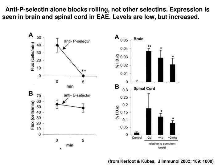
Anti-P-selectin alone blocks rolling, not other selectins. Expression is seen in brain and spinal cord in EAE. Levels are low, but increased. (from Kerfoot & Kubes, J Immunol 2002; 169: 1000)
P-selectin blocking before assay inhibits both rolling and adhesion at peak disease (4d) a4 blockade inhibits adhesion more than rolling Since rolling precedes adhesion, then P-selectin seems more important that a4 integrins (from Kerfoot & Kubes, J Immunol 2002; 169: 1000)
Both memory and effector CD4+ T cells express CD45RO - how to distinguish between the two? Ans. CCR7 CCR7- CD45RO+ T cells are effector-memory T cells - make cytokines, perforin, granzymes These cells don’t enter lymph nodes ’Combination code' for entry of T cells to normal lymph nodes is L-selectin binding, followed by CCR7 signaling through SLC. This code is expressed by naive T cells and CCR7+ memory T cells, but not by certain cells that do not recirculate, and memory-effector T cells Other effector-memory T cells: CD8+ CD11b+ cells
DC enter LN via afferent lymph -> subcapsular sinus -> T cell areas SLC and ELC are involved in this migration Maturing DC express chemokines that can attract T cells Antigen regulates microenviromental homing Integrin activation Chemokine receptor expression CCR7low CXCR5+ activated Th2 T cells may be favoured for T:B interaction - move towards and into B follicles for GC reaction Adhesion signaling also regulates microenviromental homing
L-selectin signaling Anti-L-selectin antibody induces Ca++ fluxes and tyrosine phosphorylation in PMN's Other selectin signaling Interaction between PMN's or monocytes with HUVEC's induced Ca++ fluxes in HUVEC's - blocked by antibodies against E-selectin or VCAM-1. Antibodies against P- or E-selectin induced rearrangement of EC microfilaments or actin stress fibers. ICAM-1 signaling Predominantly expressed on inflamed endothelia or on APC’s High affinity integrins bind ICAM-1 ICAM-1 induces endothelial cells to make TNF Induces astrocytes (brain), synovial cells (joints) and endothelial cells to make IL-1 and IL-6 Induces cell cycle arrest in B cells - MAPK, blocks NFkB,
Integrins a/ß heterodimers 17 a’s, 8 ß’s, making 22 integrin receptors Bind to specific receptors (ICAM’s), or to ECM (fibronectin, collagen) LFA-1 (CD11a/CD18, or aL:ß2) Mac-1 (CD11b/CD18, or aM:ß2) VLA-4 (a4:ß1) Many integrins recognize RGD motifs (arg-gly-asp) in ECM Integrin ligand-binding activity regulated by intracellular signaling (‘inside-out signaling’) Integrins associate with cytoskeleton (eg. Talin) when cells are activated.
Integrin signaling • FAK (focal adhesion kinase) • Integrin-associated tyrosine kinase, involved in integrin signaling for cytoskeletal reorganisation. • GTP-binding proteins (Rho, Ras and Rac), MAPKinase. • There are also FAK- and Ras-independent pathways • Outcome of integrin signaling: • prevention of apoptosis (stabilisation of Bcl-2 expression) • cell cycle arrest/progression • local regulation of adhesion, contractility and cell movement • Shape change • Stress fibers, and actin • modulation of growth factor signaling
b a Ras Integrin signaling 101 TCR/CD3 Ras FAK PI-3-K PIP-5-K Rho Rac PIP2 Rho-K MAPK Profilin Gelsolin Vinculin Myosin phosphatase Survival Proliferation Focal adhesions Stress fibers (from Kumar, CC. 1998. Oncogene 17:1365)
Kim et al made constructs of aL and ß2 (aL/ß2 integrin = LFA-1) coupled to mCFP and mYFP respectively and used FRET to measure interaction between the two chains. • Results show conformational changes in integrin cytoplasmic domains associated with activation in living cells. • Activation of conformational change by PMA (‘inside-out’ signaling) resulted in physical separation of cytoplasmic regions. • Ligand binding (sICAM-1) also induced spatial separation of cytoplasmic domains • Not all activation stimuli did this (eg. Mn++) • Enforced proximity of cytoplasmic regions prevented activation • GFKKR motif in a-chain transmembrane/cytoplasmic region critical for spatial separation • Talin binding to LFA-1 induces spatial separation • Chemokine signaling (SDF-1/CXCL12 to CXCR4) (‘inside-out’) also induced spatial separation
Kim, M., Carman, C.V., Springer, T.A. 2003. Science 301:1720-1725
Kim, M., Carman, C.V., Springer, T.A. 2003. Science 301:1720-1725