Download
1 / 1
10 likes | 183 Views
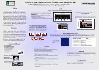
NHS. Cambridge University. CRUK-CRI. Linking tissue microarray core image data with clinical data. IIS. TMA images and meta-data. Ariol Web Service. Java Client. slidemap. Analysis results. Strangeways Laboratory. TMAR. Excel Map.xml. TMA Results.

E N D
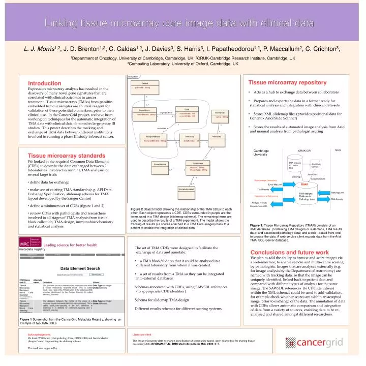
NHS Cambridge University CRUK-CRI Linking tissue microarray core image data with clinical data IIS TMA images and meta-data Ariol Web Service Java Client slidemap Analysis results Strangeways Laboratory TMAR Excel Map.xml TMA Results L. J. Morris1,2, J. D. Brenton1,2, C. Caldas1,2, J. Davies3, S. Harris3, I. Papatheodorou1,2, P. Maccallum2, C. Crichton3, 1Department of Oncology, University of Cambridge, Cambridge, UK; 2CRUK-Cambridge Research Institute, Cambridge, UK 3Computing Laboratory, University of Oxford, Cambridge, UK Pathology.xml TMA designs TMA results Pathology data Department of Astronomy TMA Results Analysis Results Images+meta-data Introduction Expression microarray analysis has resulted in the discovery of many novel gene signatures that are correlated with clinical outcomes in cancer treatment. Tissue microarrays (TMAs) from paraffin-embedded tumour samples are an ideal reagent for validation of these potential biomarkers, prior to their clinical use. In the CancerGrid project, we have been working on techniques for the automatic integration of TMA data with clinical data obtained in large phase III studies. This poster describes the tracking and exchange of TMA data between different institutions involved in running a phase III study in breast cancer. • Tissue microarray repository • Acts as a hub to exchange data between collaborators • Prepares and exports the data in a format ready for statistical analysis and integration with clinical data-sets • Stores XML slidemap files (provides positional data for GenentixAriol Slide Scanner) • Stores the results of automated image analysis from Ariol and manual analysis from pathologist scoring • Tissue microarray standards • We looked at the required Common Data Elements (CDEs) to describe the data exchanged between 2 laboratories involved in running TMA analysis for several large trials • define data for exchange • make use of existing TMA standards (e.g. API Data Exchange Specification, slidemap schema for TMA layout developed by the Sanger Centre) • define a minimum set of CDEs (figure 1 and 2) • review CDEs with pathologists and researchers involved in all stages of TMA analysis from tissue block collection, TMA design, immunohistochemistry and statistical analysis Figure 2Object model showing the relationship of the TMA CDEs to each other. Each object represents a CDE. CDEs surrounded in purple are the terms used in a TMA design (slidemap schema). The remaining terms are used to describe the results of a TMA experiment. The model allows the tracking of results (i.e scores attached to a TMA Core images) back to a patient to enable the integration of clinical data. Figure 3. Tissue Microarray Repository (TMAR) consists of an XML database (containing TMA designs or slidemaps, TMA results data; and associated pathology data) and a web –based front-end to browse the data. A web service client exports data from the Ariol TMA SQL-Server database. • The set of TMA CDEs were designed to facilitate the exchange of data and annotate: • a TMA block/slide so that it could be analysed in a different laboratory from where it was created. • a set of results from a TMA so they can be integrated into external databases • Schemas annotated with CDEs, using SAWSDL references (to appropriate CDE identifier) • Schema for slidemap TMA design • Different results schemas for different scoring systems Conclusions and future work We plan to add the ability to browse and score images via a web-interface, to enable remote and multi-centre scoring by pathologists. Images that are analysed externally (e.g. for image analysis by the Department of Astronomy) are named with tracking data, so that the image can be uniquely identified, linked back to patient data and compared with different types of analysis for the same image. The SAWSDL references (to CDE identifiers) within the XML schemas could be used to add validation, for example check whether scores are within an accepted range, prior to exchange of the data. The annotation of data with CDEs allows automatic comparison and integration of data from a variety of sources, enabling data to be re-analysed and shared amongst different researchers. C Figure 1Screenshot from the CancerGrid Metadata Registry, showing an example of two TMA CDEs Literature cited The tissue microarray data exchange specification: A community-based, open source tool for sharing tissue microarray data Berman et al, BMC Med Inform DecisMak. 2003; 3: 5. Acknowledgments We thank Will Howat (Histopathology Core, CRUK-CRI) and Gareth Maslen (Sanger Centre) for providing the slidemap schema. This work was supported by…