Download
1 / 1
10 likes | 142 Views
Severity of Disease, Radiographic Presentation, and Outcomes in Ten Adult Patients with Mycoplasma pneumoniae Pneumonia: Results from the Rapid Empiric Treatment with Oseltamivir Study (RETOS ) Cynthia Meza¹, Fabiola Gianella¹, Sriram Maranganti¹, Amy Holloway¹, Raul Nakamatsu¹ ׳ ²

E N D
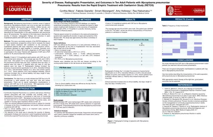
Severity of Disease, Radiographic Presentation, and Outcomes in Ten Adult Patients with Mycoplasma pneumoniae Pneumonia: Results from the Rapid Empiric Treatment with Oseltamivir Study (RETOS) Cynthia Meza¹, FabiolaGianella¹, SriramMaranganti¹, Amy Holloway¹, Raul Nakamatsu¹׳² 1. Division of Infectious Diseases, University of Louisville, 2. Division of Infectious Diseases, Robley Rex VA Medical Center RESULTS (Cont’d) ABSTRACT MATERIALS AND METHODS RESULTS • A total of 10 hospitalized patients with CAP due to Mycoplasma pneumoniae were analyzed. • The average age was 46 and 90% were male. One patient was admitted to the ICU. The table 1 shows the clinical characteristics of Pneumonia patients on admission to hospital. Study design and Study population • This was a secondary analysis of RETOS database, an ongoing, randomized, prospective clinical trial to evaluate the impact of rapid empiric treatment with Oseltamivir on the outcomes of hospitalized patients with LRTIs, in 4 hospitals in Louisville, Kentucky, during 2010/2012 influenza season. • Patients signed consent for enrollment in the study and answered a questionnaire regarding the duration of signs and symptoms of the disease with. The study was approved by the Ethics Committee of each hospital. Study definitions CAP was defined as the presence of a new pulmonary infiltrate on a chest radiograph at the time of hospitalization that was associated with at least one of the following: • New or increased cough • An abnormal temperature (<35.6˚C or > 37˚C) • Leukocytosis(leukocyte count >10,500 cells/µL),leucopenia (leukocyte count < 4,500 cells/µL), or left shift (>5% immature neutrophils) • RT- PCR (+) for Mycoplasma pneumoniae Background: Mycoplasma pneumoniae a common cause of atypical pneumonia. Mycoplasma infection can occur at any age, but infection rates are highest among school aged children, military recruits, and college students. M. pneumoniaeis considered a mild form of community-acquired pneumonia(CAP). There is little literature describing the characteristics of the adult population with pneumonia due to M. pneumoniae. The objective of the study was to describe the severity of disease, radiographic findings and outcomes in adult hospitalized patients with CAP due to M. pneumoniae. Methods: This was a secondary analysis of the RETOS database, an ongoing, randomized, prospective clinical trial to evaluate the impact of rapid empiric treatment with oseltamivir on the outcomes of hospitalized patients with lower respiratory tract infections (LRTIs). All patients admitted to eight hospitals in Louisville, Kentucky from December 2010 to March 2012 with a diagnosis of LRTI were invited to participate in the study. Patients with diagnosis of CAP and a PCR positive for M. pneumoniaewere included. Results: A total of 10 hospitalized adult patients with CAP due to M. pneumoniae were analyzed. The average age was 46, and 9 (90%) were male. One patient (10 %) was admitted to the ICU. Based on the pneumonia severity index risk class, 2 patients were risk class I, 4 patients were risk class II, 2 patients were risk class III, and two patients were risk class IV. Radiographic findings included: the presence of parenchymal opacification (n=4, 40%), interstitial infiltrate (n=3, 30%), reticulonodular infiltrate (n=2, 20%) and nodular opacity (n=1, 10%). Seven patients (70%) had multilobar infiltrates. Clinical outcomes included: time to clinical stability, 4±2 days; length of stay, 6±2 days; mortality (0, 0%). Conclusions: The data from our study indicate that CAP due to by M. pneumoniae in adult patients can lead both mild and severe presentation. There are no typical radiographic manifestations in patients with CAP due to M. pneumoniae. With appropriate empiric therapy, good patient outcomes can be expected. . • . Table 2: Frequency of lobe involvement. CONCLUSIONS Patients were classified into five PSI risk classes according to the scoring system based on the risk factors as shown below: Table 1 : Our study indicates that CAP due to Mycoplasma pneumoniae in adult patients can lead both mild and severe presentation. There are not typical radiographic manifestations in patients with Cap due to Mycoplasma pneumoniae Very few studiesdescribing the characteristics of the adult population with pneumonia due to Mycoplasma pneumoniae With appropriate empiric therapy at the right time, good patient outcomes can be expected. Radiographic findings (fig 1) included: the presence of parenchymal opacification (n=4, 40%), interstitial infiltrate (n=3,30%),reticulonodular infiltrate (n=2,20%) and nodular opacity (n=1,10%). Seven patients have multilobar infiltrate (table 2). Patients were treated empirically with macrolide. Clinical outcomes included: time to clinical stability, 4±2 days; length of stay, 6±2 days; mortality (0,0%). Time to clinical stability criteria was defined as the day that the following four criteria were met REFERENCES • Fahmi S, BibianaC, OrnaN, et al. Etiology of Community-Acquired Pneumonia in Hospitalized Patients in Northern Israel. IMAJ • VOL 12 • August 2010 • 2. MonyS, Moshe R-A, Uzi I, et. al.Massiveempyema caused by Mycoplasma pneumoniaein an adult: A case report. BMC Infectious Diseases 2006, 6:18 doi:10.1186/1471-2334-6-18. • 3. Mandell LA, Wunderink RG, Anzueto A, et al. Infectious Diseases Society of America/American Thoracic Society consensus guidelines on the management of community-acquired pneumonia in adults. Clin Infect Dis 2007; 44(suppl):S27-S72 • 4. Naoyuki M, Tadaaki S, Yasuhiro K, et al. Radiographicfeatures of Mycoplasmapneumoniaepneumonia: differential diagnosis and performance timing. BMC Medical Imaging 2009, 9:7 doi:10.1186/1471-2342/9/7 • 5. Stephen G. Mycoplasmapneumoniaeinfection in adults. www.uptodate.com INTRODUCTION • Community Acquired Pneumonia is a common and serious infectious disease associated with high morbidity and mortality rates. (1) Mycoplasma Pneumoniae is considered a mild form of Community acquired pneumonia (CAP). Mycoplasma pneumoniae is responsible for more than 20 % of Community Acquired Pneumonia cases, and capable of causing upper respiratory illness as well. (2) • There are several published pneumonia scores to assess for severity of disease at the time of hospitalization and predict clinical outcomes in patients with CAP [3]. • Time to clinical stability (TCS) is an important early outcome in hospitalized patients with CAP. • The objective of the study was to describe the severity of disease, radiographic findings and outcomes in adult hospitalized patients with Community acquired Pneumonia due to Mycoplasma pneumoniae. Study sample Nasopharyngeal (NP) and oropharyngeal (OP) swabs were collected at the time of study entry. OP swabs were used for detection of Legionella pneumophila, Chlamydophila pneumonia and Mycoplasma pneumoniae. RT-PCR was used for detection. • Statistical Analysis • For patients with Mycoplasma pneumoniae, frequencies and percentages were used to describe categorical variables and means were used to describe continuous variables. . Figure 1. Radiographic findings of patyeints with Mycoplasma pneumoniae