Download
1 / 29
300 likes | 535 Views
Aims. Introduction to the heart. Heart contraction and electrical conduction. Readings; Sherwood, Chapter 9. Cardiovascular Physiology. Cardiac Muscle Myocytes (cardiac muscle cells) Myocytes are connected to each other via __Intercalated Disks__

E N D
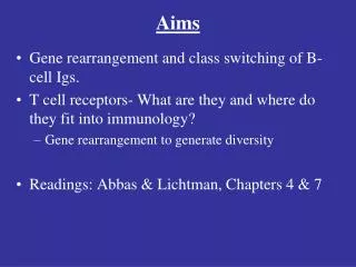
Aims • Introduction to the heart. • Heart contraction and electrical conduction. • Readings; Sherwood, Chapter 9
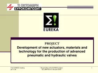
Cardiovascular Physiology • Cardiac Muscle • Myocytes (cardiac muscle cells) • Myocytes are connected to each other via __Intercalated Disks__ • Composed of Desmosomes and Gap Junctions • Allow waves of action potentials to spread from one cell to the next (Syncytium). Sherwood’s Human Physiology 9-8 (9-6 6th Edition)
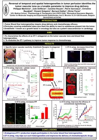
Human Heart Anatomy Sherwood’s Human Physiology 9-5 (9-4 6th Edition)
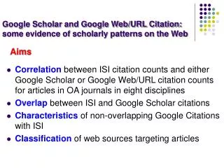
Valve Physiology • Valves are mechanical devices and function in response to blood flow. • They function on the principle that they stay open as long as the blood pressure is greater in the emptying chamber. • They close as soon as the blood pressure is greater in the filling chamber, thus blood flow is unidirectional. P1 P2 P1 P2 Sherwood’s Human Physiology 9-4 (9-3 6th Edition)
Ion Concentrations • Inside a cell • K+ • Outside a cell • Na + • Ca + • Cl- Guyton’s Textbook of Medical Physiology 4-1
Cell Membrane Transport • Passive • Simple diffusion • Facilitated diffusion • Active (needs energy) Guyton’s Textbook of Medical Physiology 4-2
Two Types of Cardiac Cells • Autorhythmic Cells • __non-contractile_________ • Initiate and conduct action potentials responsible for contraction. • Located in the SA node, AV node, Bundle of His, Purkinje fibers. • Contractile Cells • 99% of the cardiac muscle cells
Specialized Conduction System • Sinoatrial (SA) node. • _Pacemaker____ • Cells exhibit autorhythmicity. Sherwood’s Human Physiology 9-11 (9-8 6th Edition)
Specialized Conduction System • Atrioventricular (AV) node • Delays electrical signal due to a decreased number of gap junctions. Sherwood’s Human Physiology 9-11 (9-8 6th Edition)
Conduction Delay • Atrioventricular fibrous tissue • Acts as an insulator Guyton’s Textbook of Medical Physiology 10-3
Specialized Conduction System • Atrioventricular (AV) bundle or Bundle of His. • Transmits electrical signal down to the ventricles. • Purkinje fibers • Send action potential through ventricles. • Has increased number of gap junctions. Sherwood’s Human Physiology 9-11 (9-8 6th Edition)
Pacemaker Potential • Decreased K+ efflux and constant Na+ influx via leak channels. • Resulting in a higher resting potential. • Slow Ca++ inward permeabilityvia transient voltage-gated Ca++ channel. Sherwood’s Human Physiology 9-10 (9-7 6th Edition)
Pacemaker Potential • Ca++ inward permeability via longer lasting voltage-gated Ca++ channel. • Resulting in __faster depolarization___ Sherwood’s Human Physiology 9-10 (9-7 6th Edition)
Pacemaker Potential • K+ outward permeability via voltage-gated channel. • Resulting in repolarization Sherwood’s Human Physiology 9-10 (9-7 6th Edition)
Pacemakers • SA node (normal pacemaker) • 70-80 action potentials per minute. • Ectopic Pacemakers • AV node • 40-60 action potentials per minute. • Bundle of His and purkinje fibers • 20-40 action potentials per minute.
Pacemakers Sherwood’s Human Physiology 9-12 5th Edition only
Abnormal Conduction Pathway • Normal • -SA node sets the pace • SA Node non-functional • -AV node sets the pace at a slower rate • AV Node non-functional • -Atria contract at SA node rate while ventricles contract at Purkinje fiber rate (much slower) • -Complete heart block that requires an artificial pacemaker. • Purkinje fiber is hyper-excitable • - Called an ectopic focus that causes a premature beat. Guyton’s Textbook of Medical Physiology 10-4 Sherwood’s Human Physiology 9-15
Abnormal Conduction Pathway Guyton’s Textbook of Medical Physiology 10-4 Sherwood’s Human Physiology 9-15
Cardiac Muscle Cell Action Potential • Depolarization • Na+ inward • Plateau • Ca++ inward • Repolarization • K+ outward Sherwood’s Human Physiology 9-15 (9-11 6th Edition)
SA node potentials vs. cardiac cell potentials • SA node has a higher resting potential than other cardiac muscle cells. Guyton’s Textbook of Medical Physiology 10-2
ECC in Cardiac Muscle • The majority of Ca++ required for contraction comes from the sarcoplasmic reticulum and not the ECF. Sherwood’s Human Physiology 9-16 (9-12 6th Edition)
Refractory Period • Long refractory period important because it makes tetanus impossible. Sherwood’s Human Physiology 9-17 (9-12 6th Edition)
Requirements for Efficient Cardiac Contraction • Atrial excitation and contraction need to be complete before ventricular contraction occurs. • Excitation of cardiac muscle fibers should be coordinated so that each chamber contracts as a unit. • Pair of atria and pair of ventricles should be coordinated so that both members of the pair contract simultaneously.
What is an Electrocardiogram (ECG or EKG)? • It is not a direct recording of the actual electrical activity of the heart. • It measures the portion of the electrical activity of the heart that is transduced in the body fluids and reaches the body surface. • It is a complex recording that represents the overall activity throughout the heart during depolarization and repolarization. • Not a single cell measurement
ECG • 6 body & 6 chest leads for a total of 12 leads Sherwood’s Human Physiology 9-18 (9-14 6th Edition)
ECG • P wave- atrial depolarization • PR segment- AV nodal delay • QRS complex- ventricular depolarization and atrial repolarization • ST segment- ventricular contraction and emptying • T wave- ventricular repolarization • TP interval- ventricular filling Sherwood’s Human Physiology 9-19 (9-15 6th Edition)
Abnormal ECG • Rate Abnormalities • Tachycardia (>100 beats/min.) • Rhythm Abnormalities (Arrhythmias) • Extrasystole (premature beat) • Ventricular fibrillation • Atrial fibrillation • Complete Block • Cardiac Myopathies • Myocardial Infarction Sherwood’s Human Physiology 9-20 (9-16 6th Edition)
Next Time • Cardiac Cycle • Cardiac regulation • Extrinsic vs. intrinsic • Reading; Sherwood, Chapter 9
Objectives • Describe the structure and function of cardiac myocytes. • Describe the anatomy of the heart and how blood flows through it. • Describe cardiac contraction • Conduction (normal and abnormal) • Pacemakers • Action potentials • Refractory period • Describe the ECG. • P Wave, QRS Complex, T Wave, Abnormal ECG