Download
1 / 1
10 likes | 85 Views
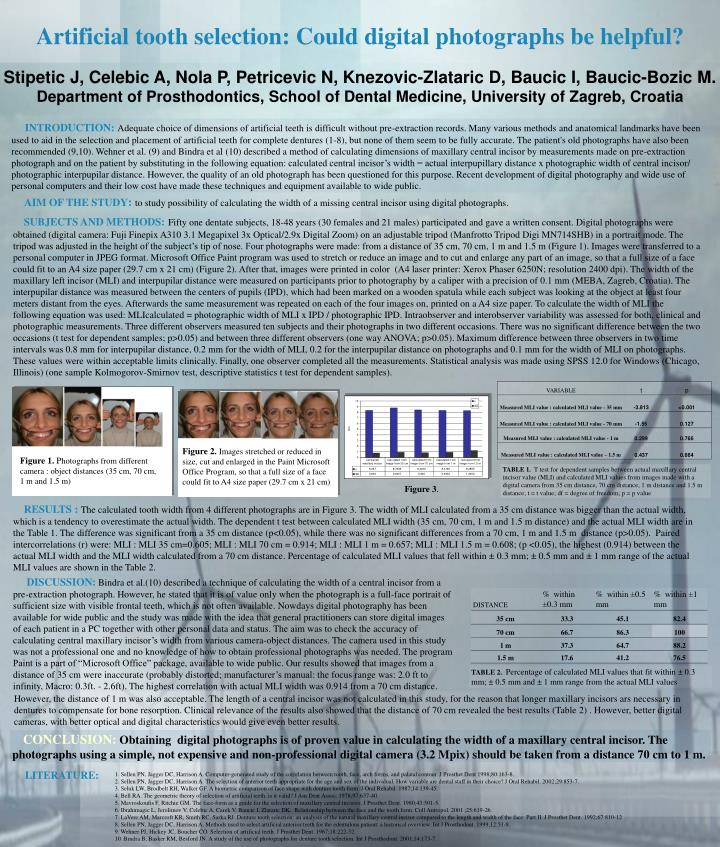
Figure 2. Images stretched or reduced in size, cut and enlarged in the Paint Microsoft Office Program, so that a full size of a face could fit to A4 size paper (29.7 cm x 21 cm). Figure 1. P hotographs from different camera : object distances (35 cm, 70 cm, 1 m and 1.5 m).

E N D
Figure 2.Images stretched or reduced in size, cut and enlarged in the Paint Microsoft Office Program, so that a full size of a face could fit to A4 size paper (29.7 cm x 21 cm) Figure 1.Photographs from different camera : object distances (35 cm, 70 cm, 1 m and 1.5 m) INTRODUCTION:Adequate choice of dimensions of artificial teeth is difficult without pre-extraction records. Many various methodsand anatomical landmarks have been used to aid in the selection and placement of artificial teeth for complete dentures (1-8), but none of them seem to be fully accurate. The patient's old photographs have also been recommended (9,10). Wehner et al. (9) and Bindra et al (10) described a method of calculating dimensions of maxillary central incisor by measurements made on pre-extraction photograph and on the patient by substituting in the following equation: calculated central incisor’s width = actual interpupillary distance x photographic width of central incisor/ photographic interpupilar distance. However, the quality of an old photograph has been questioned for this purpose. Recent development of digital photography and wide use of personal computers and their low cost have made these techniques and equipment available to wide public. Artificial tooth selection: Could digital photographs be helpful? AIM OF THE STUDY:to study possibility of calculating the width of a missing central incisor using digital photographs. SUBJECTS AND METHODS:Fifty one dentate subjects, 18-48 years (30 females and 21 males) participated and gave a written consent.Digital photographs were obtained (digital camera: Fuji Finepix A310 3.1 Megapixel 3x Optical/2.9x Digital Zoom) on an adjustable tripod (Manfrotto Tripod Digi MN714SHB) in a portrait mode. The tripod was adjusted in the height of the subject’s tip of nose. Four photographs were made: from a distance of 35 cm, 70 cm, 1 m and 1.5 m (Figure 1). Images were transferred to a personal computer in JPEG format. Microsoft Office Paint program was used to stretch or reduce an image and to cut and enlarge any part of an image, so that a full size of a face could fit to an A4 size paper (29.7 cm x 21 cm) (Figure 2). After that, images were printed in color (A4 laser printer: Xerox Phaser 6250N; resolution 2400 dpi). The width of the maxillary left incisor (MLI) and interpupilar distance were measured on participants prior to photography by a caliper with a precision of 0.1 mm(MEBA, Zagreb, Croatia). The interpupilar distance was measured between the centers of pupils (IPD), which had been marked on a wooden spatula while each subject was looking at the object at least four meters distant from the eyes. Afterwards the same measurement was repeated on each of the four images on, printed on a A4 size paper. To calculate the width of MLI the following equation was used: MLIcalculated = photographic width of MLI x IPD / photographic IPD.Intraobserver and interobserver variability was assessed for both, clinical and photographic measurements. Three different observers measured ten subjects and their photographs in two different occasions. There was no significant difference between the two occasions (t test for dependent samples; p>0.05) and between three different observers (one way ANOVA; p>0.05). Maximum difference between three observers in two time intervals was 0.8 mm for interpupilar distance, 0.2 mm for the width of MLI, 0.2 for the interpupilar distance on photographs and 0.1 mm for the width of MLI on photographs. These values were within acceptable limits clinically. Finally, one observer completed all the measurements.Statistical analysis was made using SPSS 12.0 for Windows (Chicago, Illinois) (one sample Kolmogorov-Smirnov test, descriptive statistics t test for dependent samples). Stipetic J, Celebic A, Nola P, Petricevic N, Knezovic-Zlataric D, Baucic I, Baucic-Bozic M. Department of Prosthodontics, School of Dental Medicine, University of Zagreb, Croatia TABLE 1. T test for dependent samples between actual maxillary central incisor value (MLI) and calculated MLI values from images made with a digital camera from 35 cm distance, 70 cm distance, 1 m distance and 1.5 m distance; t = t value; df = degree of freedom; p = p value Figure 3. RESULTS :The calculated tooth width from 4 different photographs are in Figure 3. The width of MLI calculated from a 35 cm distance was bigger than the actual width, which is a tendency to overestimate the actual width. The dependent t test between calculated MLI width (35 cm, 70 cm, 1 m and 1.5 m distance) and the actual MLI width are in the Table 1. The difference was significant from a 35 cm distance (p<0.05), while there was no significant differences from a 70 cm, 1 m and 1.5 m distance (p>0.05).Paired intercorrelations (r) were: MLI : MLI 35 cm=0.605; MLI : MLI 70 cm = 0.914; MLI : MLI 1 m = 0.657; MLI : MLI 1.5 m = 0.608; (p <0.05), the highest (0.914) between the actual MLI width and the MLI width calculated from a 70 cm distance.Percentage of calculated MLI values that fell within ± 0.3 mm; ± 0.5 mm and ± 1 mm range of the actual MLI values are shown in the Table 2. DISCUSSION:Bindra et al.(10) described a technique of calculating the width of a central incisor from a pre-extraction photograph. However, he stated that it is of value only when the photograph is a full-face portrait of sufficient size with visible frontal teeth, which is not often available. Nowdays digital photography has been available for wide public and the study was made with the idea that general practitioners can storedigitalimages of each patient in a PC together with other personal data and status. The aim was to check the accuracy of calculating central maxillary incisor’s width from various camera-object distances. The camera used in this study was not a professional one and no knowledge of how to obtain professional photographs was needed. The program Paint is a part of “Microsoft Office” package, available to wide public. Our results showed that images from a distance of 35 cm were inaccurate(probably distorted; manufacturer’s manual: the focus range was: 2.0 ft to infinity, Macro: 0.3ft. - 2.6ft). The highest correlation with actual MLI width was 0.914 from a 70 cm distance. TABLE 2.Percentage of calculated MLI values that fit within ± 0.3 mm; ± 0.5 mm and ± 1 mm range from the actual MLI values However, the distance of 1 m was also acceptable. The length of a central incisor was not calculated in this study, for the reason that longer maxillary incisorsars necessary in dentures to compensate for bone resorption. Clinical relevance of the results also showed that the distance of 70 cm revealed the best results (Table 2) . However, better digital cameras, with better optical and digital characteristics would give even better results. CONCLUSION:Obtaining digital photographs is of proven value in calculating the width of a maxillary central incisor. The photographs using a simple, not expensive and non-professional digital camera (3.2 Mpix) should be taken from a distance 70 cm to 1 m. LITERATURE: 1. Sellen PN, Jagger DC, Harrison A. Computer-generated study of the correlation between tooth, face, arch forms, and palatal contour. J Prosthet Dent 1998;80:163-8. 2. Sellen PN, Jagger DC, Harrison A. The selection of anterior teeth appropriate for the age and sex of the individual. How variable are dental staff in their choice? J Oral Rehabil. 2002;29:853-7. 3. Seluk LW, Brodbelt RH, Walker GF. A biometric comparison of face shape with denture tooth form. J Oral Rehabil. 1987;14:139-45. 4. Bell RA. The geometric theory of selection of artificial teeth: is it valid? J Am Dent Assoc. 1978;97:637-40. 5. Mavroskoufis F, Ritchie GM. The face-form as a guide for the selection of maxillary central incisors. J Prosthet Dent. 1980;43:501-5. 6. Ibrahimagic L, Jerolimov V, Celebic A, Carek V, Baucic I, Zlataric DK. Relationship between the face and the tooth form. Coll Antropol. 2001 ;25:619-26. 7. LaVere AM, Marcroft KR, Smith RC, Sarka RJ. Denture tooth selection: an analysis of the natural maxillary central incisor compared to the length and width of the face: Part II. J Prosthet Dent. 1992;67:810-12 8. Sellen PN, Jagger DC, Harrison A. Methods used to select artificial anterior teeth for the edentulous patient: a historical overview. Int J Prosthodont. 1999;12:51-8. 9. Wehner PJ, Hickey JC, Boucher CO. Selection of artificial teeth. J Prosthet Dent. 1967;18:222-32. 10. Bindra B, Basker RM, Besford JN. A study of the use of photographs for denture tooth selection. Int J Prosthodont. 2001;14:173-7.