Download
1 / 24
240 likes | 382 Views
01/26/07. How NMR is Used for the Study of Biomacromolecules. Analytical biochemistry Comparative analysis Interactions between biomolecules Structure determination Biomolecular dynamics from NMR. Arunkumar et al., JBC 278 , 41077-41082 (2003) Mer et al. Cell 103 , 449-456 (2000)

E N D
01/26/07 How NMR is Used forthe Study of Biomacromolecules • Analytical biochemistry • Comparative analysis • Interactions between biomolecules • Structure determination • Biomolecular dynamics from NMR Arunkumar et al., JBC 278, 41077-41082 (2003) Mer et al. Cell 103, 449-456 (2000) Ohi et al. NSB 11, 250-255 (2003)
Analytical Protein Biochemistry • Purity (can detect >99%)- heterogeneity, degradation, contamination • Is a protein structured?- fast and easy assay, detects aggregation and folding • Check on sequence (fingerprint regions) • Don’t need the sequence-specific assignments!
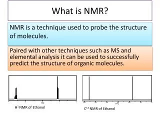
Protein Folding and Fingerprints 1H COSY 15N-1H HSQC 13C HSQC also! Assay of tertiary structure & check sequence
Comparative Analysis • Different preparations, changes in conditions • Domain structure • Structural heterogeneity (e.g. Pro cis-trans isomerization) • Homologous proteins, mutants, engineered proteins
B A B A RPA70 15N 15N 15N 2 2 3 1H 3 1 1 1H 1H Arunkumar et al., JBC (2003) Folding and Domain StructureAre domains packed together or independent? • Chemical shift is extremely sensitive • If peaks are the same, structure is the same • If peaks are different, the structure is different but we don’t know how much
Biochemical Effect of MutationsAssay for proper folding/stability Wild-type Partially destabilized Structural heterogeneity Unfolded Ohi et al., NSB (2003)
Structural Basis for PhenotypeWhat is the cause of defective RNA splicing by Prp19-1? Initial interpretation was defect in some binding interface NMR showed U-box folding defect Ohi et al., NSB (2003)
NMR to Study Interactions • Monitor the binding of molecules • Determine binding constants (discrete off rates, on rates) • Sequence and 3D structural mapping of binding interfaces
NMR- The Master Spectroscopy Titration monitored by 15N-1H HSQC NMR Provides • Site-specific • Multiple probes • In-depth information • Perturbations can be mapped on structure
Binding Constants FromChemical Shift Changes Stronger Weaker Molar ratio of d-CTTCA • Fit change in chemical shift to binding equation Arunkumar et al., JBC (2003)
Probe Binding Events by NMR15N-RPA32C + Unlabeled XPA1-98 15N-1H HSQC • Only 19 residues affected • Discrete binding site • Signal broadening exchange between the bound and un-bound state • Kd ~ 1 mM RPA32C RPA32C + XPA 1-98 Mer et al., Cell (2000)
C N Map XPA Binding Site on RPA32C Using NMR • Map chemical shift perturbations on the structure of RPA32C • Can even map directly on to sequence with no structure Mer et al., Cell (2000)
NMR Experimental Observables Providing Structural Information • Distances from dipolar couplings (NOEs) • Backbone and side chain dihedral angles from scalar couplings • Backbone conformation from chemical shifts (Chemical Shift Index- CSI): , • Hydrogen bonds- NH exchange or J • Relative inter-nuclear orientations from residual dipolar couplings (RDCs)
NMR Structure Calculations • Objective is to determine all conformations consistent with the experimental data • Programs initially search geometry only • More calculations using molecular force fields to improve molecular properties • NMR data are not perfect (noise, incomplete) multiple solutions (conformational ensemble)
Characteristics of NMR Structures • Secondary structures well defined, loops variable • Interiors well defined, surfaces more variable • RMSD provides measure of variability
Restraints and Uncertainty • Large # of restraints = low values of RMSD • The most important restraints are long-range
Assessing the Accuracy and Precisionof NMR Structures • Number of experimental restraints (A/P) • Violation of constraints- number, magnitude (A) • Compare model and exptl. parameters (A) • Comparison to known structures: PROCHECK (A) • Molecular energies (?A?, subjective) • RMSD of structural ensemble (P, biased)
Biomolecular Dynamics from NMR Why? Function requires motion/kinetic energy • Entropic contributions to binding events • Protein folding/unfolding • Uncertainty in NMR and crystal structures • Effect on NMR experiments dynamics to predict outcomes and design new experiments • Calibration of computational methods
Characterizing Protein Dynamics: Parameters/Timescales Residual Dipolar Couplings
NMR Observables and Dynamics • Number of signals per atom: multiple signals for slow exchange between conformational states • Linewidths: narrow = faster motion, wide = slower; dependent on MW and conformational states • Exchange of NH with solvent:slow timescales • NMR relaxation measurements (ps-ns, ms-ms) • Direct measurements of motion of atoms • Parameters: R1 (T1), R2 (T2), Het. NOE (e.g. 15N- 1H)
B A B A 15N 15N 15N 1H 1H 1H Linewidth is Dependent on MW • Linewidth determined by size of particle • Fragments have narrower linewidths Arunkumar et al., JBC (2003)
40 173 P Independent Domains in Large Proteins RPA32 RPA14 > 400 residues / ~80 signals Why? A structurally-independent functional domain
Correlating Structure and Dynamics Weak correlation • Measurements show if high RMSD is due to high flexibility (low S2) Strong correlation