Download
1 / 32
1.5k likes | 7.95k Views
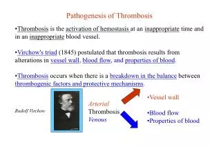
•Vessel wall •Blood flow •Properties of blood. Arterial Thrombosis Venous. Rudolf Virchow. Pathogenesis of Thrombosis. • Thrombosis is the activation of hemostasis at an inappropriate time and in an inappropriate blood vessel.

E N D
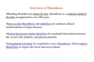
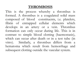
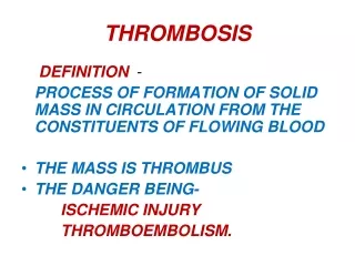
•Vessel wall •Blood flow •Properties of blood Arterial Thrombosis Venous Rudolf Virchow Pathogenesis of Thrombosis •Thrombosis is the activation of hemostasis at an inappropriate time and in an inappropriate blood vessel. •Virchow's triad (1845) postulated that thrombosis results from alterations in vessel wall, blood flow, and properties of blood. •Thrombosis occurs when there is a breakdown in the balance between thrombogenic factors and protective mechanisms.
Pathogenesis of Thrombosis (cont’d) The protective mechanisms are: 1. The nonthrombogenic properties of intact endothelium; 2. Neutralization of activated coagulation factors by endothelial cell-bound components; 3. Neutralization of activated coagulation factors by naturally occurring protease inhibitors; 4. Dilution of activated coagulation factors and disruption of platelet aggregates by blood flow; 5. Clearance of activated coagulation factors by the liver; 6. Dissolution of fibrin thrombi by the fibrinolytic system.
Overview of Approach to Treatment •The optimal approach to the prevention and treatment of thromboembolic disease differs depending on whether the process involves thevenous or arterial circulation. •The most effective way to prevent arterial thrombosis is to prevent the underlying atherosclerosis. •Complications of atherosclerosis (myocardial infarction, stroke and peripheral arterial ischemia) can be reduced: --by not smoking, --by treating hypertension, --by reducing cholesterol, and --by increasing physical activity.
Overview of Approach to Treatment (cont’d) •Thrombotic complications of coronary atherosclerosis and cerebral arteriosclerosis can be reduced by antiplatelet drugs (i.e., aspirin). •Thrombolytic therapy has proven to be very successful in lysing acute coronary thrombi and has had a dramatic effect in reducing mortality after acute myocardial infarction (time to treatment is important).
Management and Prevention •Antithrombotic medication now forms an important part of the management and prevention of: -ischemic heart disease (IHD); -stroke; -and other vascular conditions. •Thrombosis is the final structural event leading to myocardial infarction or sudden coronary death. •Reducing thrombotic potential is an effective way of preventing the onset or recurrence of clinical events regardless of risk factors.
Point of intervention Smoking Blood pressure Cholesterol Obesity Other factors Risk Thrombosis Heart attack Management and Prevention (cont’d)
Atherosclerosis •No disease in the U.S. is responsible for more deaths and has engendered more controversy than atherosclerosis (AS). •Characterized by plaques called atheromasthat protrude into the lumen, weaken the underlying media, and undergo a series of complications that predispose to overlying thrombosis. •Coronary atherosclerosis induces ischemic heart disease (IHD) and when lesions are complicated by thrombosis, myocardial infarction (MI) accounts for many deaths in the U.S.
Atherosclerosis (cont’d) •Atherothrombotic disease of the cerebral vessels is the major cause of brain infarcts, so-called strokes. •Atherosclerosis often produces ischemia of the intestines and lower extremities, and it is a major cause of aortic aneurysms that can rupture to produce fatal hemorrhage.
Risk Factors •Prevalence and severity of the disease are related to many factors, some constitutional but others are acquired and potentially controllable. •Constitutional factors are age, sex, and genetics: Age. Clinically significant disease rises with each decade, even into advanced age. Sex.Males are much more prone to AS than females. Females are sheltered from disease from AS until menopause. Genetics. Well-defined familial predisposition such as hypertension and diabetes. Genetic derangements in lipoprotein metabolism, prototype is familial hypercholesterolemia.
Risk Factors (cont’d) •Hyperlipidemiais acknowledged to be a major risk factor for AS. Most evidence implicates hypercholesterolemia. •Inverse relationship between symptomatic AS and high-density lipoprotein (HDL or HDL-C) levels. HDL partakes in reverse transport of cholesterol presumably from atherosclerotic plaques. •Hypertension is a major risk factor for atherosclerosis and may well be more important than hypercholesterolemia after age 45. •Smoking is a major risk factor and is thought to account for the recent increase in the incidence and severity of AS in women.
Risk Factors (cont’d) •Diabetes mellitus induces hypercholesterolemia. There is an increased risk of strokes and atherosclerosis-induced gangrene of the lower extremities. •Multiple factors impose more than an additive effect (hyperlipidemia, hypertension, and smoking); heart attack rate is seven times greater than when none were present. •AS may develop without apparent risk factors, so those living "the prudent life" are not make immune to this killer disease.
Atherosclerosis Definition- •Atherosclerosis is a process that impairs blood flow to various organs. •Atherosclerosis is from the Greek words, refers to the thickening of the arterial intima (sclerosis, “hardening”) and accumulation of lipid (athere, “gruel”) that characterize the typical lesion. •Atherosclerosis narrows arteries, a process that causes ischemia, infarction, and necrosis.
Atherosclerosis (cont’d) •It is a disease of the arterial intima leading to the formation of fibrous (“atheromatous”) plaques- proliferation of smooth muscle cells and the accumulation of lipids. •Atherosclerosis affects large and medium-sized arteries.
Atherosclerosis--Basic Mechanisms •Atherosclerosis is the principle source of: --cerebral and myocardial infarction, --gangrene of the extremities, and --loss of function of organs and/or tissues. •Atherosclerosis appears to be a chronic inflammatory condition that is converted to an acute clinical event by the induction of plaque rupture, which in turns leads to thrombosis and death.
Thrombus nearly occluding the lumen of the carotid artery
Atherosclerosis--Basic Mechanisms (cont’d) •This suggests that lesions of atherosclerosis represent a specialized form of a protective, inflammatory (“fibroproliferative”) response to forms of insult to the artery wall. •Depending upon the nature and duration of the insult, the protective response may become excessive and over many years in its excess become a disease process. •This is called the “response-to-injury hypothesis”.
“Response-to-Injury Hypothesis” for Development of Atherosclerosis 1. Numerous forms of insult to the vessel occur to cells of the artery cell wall. 2. Chronic inflammatory response featuring monocytes and T-lymphocytes. 3. These cells adhere to the endothelium and invade the artery wall.
“Response-to-Injury Hypothesis” for Development of Atherosclerosis 4. Many of the monocytes mature to macrophages. 5.Cytokines and growth factors, affect some of the T-cells to undergo replication. 6. Results in smooth muscle cell migration and proliferation within the intima.
“Response-to-Injury Hypothesis” for Development of Atherosclerosis 7. These events culminate in the lesions that represent different stages of this specialized inflammatory, fibroproliferative response; and these cells contain varying amounts of associated lipid and lipoproteins.
Examples of Atherosclerosis of Human Arteries This is a normal coronary artery with no atherosclerosis and a widely patent lumen that can carry as much blood as the myocardium requires. Fatty streaks in a relatively normal adult aorta. Coronary artery with mild atherosclerosis and some fatty streaks.
Examples of Atherosclerosis of Human Arteries The degree of atherosclerosis is much greater in this coronary artery, and the lumen is narrowed by half. A small area of calcification is seen in the plaque at the right. A coronary thrombosis is seen microscopically occluding the remaining small lumen of this coronary artery.
Examples of Atherosclerosis of Human Arteries Three aortas are shown to demonstrate mild, moderate, and severe atherosclerosis from bottom to top. Here is occlusive coronary atherosclerosis. The coronary at the left is narrowed by 60 to 70%. The coronary at the right is even worse with evidence for previous thrombosis with organization of the thrombus and recanalization such that there are three small lumens remaining.
Examples of Atherosclerosis of Human Arteries Microscopically, the aortic atheromatous plaque is thicker than the remaining media at the right. The plaque contains amorphous pink material with slit-like "cholesterol clefts" of lipid material. There is overlying recent hemorrhage at the left. Thrombus may form on top of such a plaque. At higher magnification, many foam cells (macrophages full of lipid material) and a cholesterol cleft are seen in this atheromatous plaque.
Mechanism of Cholesterol-lowering Drugs •Cholesterol is generally thought to be the major contributor to atherosclerosis. •If we have these deposits of cholesterol, the lipoproteins are modified, and so forth and so forth. •Lowering cholesterol, especially LDL-C (so called “bad cholesterol”), is clearly a good thing. •Lowering LDL-C helps prevent and may even reverse atherosclerosis, and it may even decrease the risk of heart attacks and strokes: -statins -bile acid-binding resins -niacin -fibrates
Statins •Inhibit 3-hydroxy-3-methylglutaryl-coenzyme A (HMG-CoA) reductase •Catalyzes conversion of HMG-CoA to mevalonate, an early step in the synthesis of cholesterol– a rate-limiting step in cholesterol production. •Inhibit that step and you inhibit your ability to make cholesterol! •Statins lower serum LDL-C concentrations by up-regulating LDL-receptor activity and by reducing entry of LDL into the circulation. •Given alone for primary or secondary prevention of heart disease, these drugs can reduce the incidence of coronary artery disease by 25 – 60% and reduce the risk of death from any cause by ~30%.
HMG-CoA (3-Hydroxy-3-methylglutaryl-CoA) Lovastatin Statins (cont’d) •One of the drugs used today is shown below next to HMG-CoA:
Lipids from diet Chylomicrons Remnants Lipoprotein lipase VLDL Peripheral cell Remnants LDL-R Cholesterol LDL LRP LDL-R Hepatic lipase Cholesterol Cholesterol-ester Transport protein Bile acids Cholesterol HDL Liver Statins (cont’d)
Lipids from diet Chylomicrons Remnants Lipoprotein lipase VLDL Peripheral cell Remnants LDL-R Cholesterol LDL LRP LDL-R Hepatic lipase Cholesterol Cholesterol-ester Transport protein Bile acids Cholesterol HDL Liver Statins (cont’d)
Statins (cont’d) Adverse effects- •The most common adverse effects of statins are gastrointestinal upset, muscle aches, and hepatitis. •Sometimes there is myopathy, rash, peripheral neuropathy, insomnia, bad or vivid dreams, and difficulty sleeping or concentrating. •Hepatotoxicity occurs in <1 % of patients. •Hepatitis — fatigue, sluggishness, anorexia, and weight loss — resemble those of an influenza-like syndrome. The symptoms subside almost overnight after the drug is discontinued. •Myotoxicity is even rarer.