Download
1 / 10
150 likes | 445 Views
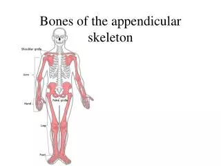
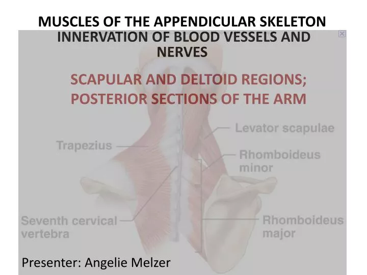
MUSCLES OF THE APPENDICULAR SKELETON INNERVATION OF BLOOD VESSELS AND NERVES. SCAPULAR AND DELTOID REGIONS; POSTERIOR SECTIONS OF THE ARM. Presenter: Angelie Melzer. MUSCLES OVERVIEW. MUSCLES OF THE BACK: ORIGIN, INSERTION AND ACTION. KEY O=Origin I=Insertion A=Action N=Nerve .

E N D
MUSCLES OF THE APPENDICULAR SKELETON INNERVATION OF BLOOD VESSELS AND NERVES SCAPULAR AND DELTOID REGIONS; POSTERIOR SECTIONS OF THE ARM Presenter: Angelie Melzer
MUSCLES OF THE BACK: ORIGIN, INSERTION AND ACTION KEY • O=Origin • I=Insertion • A=Action • N=Nerve Biomechanical actions of the shoulder: Flexion, Extension, Abduction, Adduction, Lateral Abduction and Adduction, Internal and External Rotation
Muscles Stabilizing and Moving the Head and Neck • Trapezuius: 3 Fibers ALL: O= Nuchal Line on the Occipital Protuberance, LigamentumNuchae and Spinous Processes of C7-T12. Upper: I=: Anteriorly on the medial border of the clavicle and posteriorly on the acromion A=Bilaterally: Extend head/neck, Unilaterally:Laterally Flex head and neck on same side, Rotate head to opposite side, Elevate Scapula, Upwardly rotate scapula Mid: I=Spine of the scapula to the spinous processes of T2 A=Adduct and stabilize the scapula Lower: I=Spine of the scapula to the spinous processes of T12 A=Depress the scapula, Upwardly rotate the scapula • N=Spinal portion of the cranial nerve XI and ventral ramus C2-4 • Synergists: Levator scapulae and Rhomboids
Muscles Stabilizing and Moving the Shoulder Girdle and Torso GLENOHUMERAL JOINT MUSCLES: • Deltoid: O=Lateral 1/3 of the clavicle, acromion and spine of the scapula I=Deltoid Tuberosity A=Shoulder Abduction, Posterior Fibers: Extend, Laterally Rotate and Horizontally Abduct the shoulders *The anterior and posterior fibers act as antagonists to each other. N=Axillary C5-6 • Rotator Cuff Muscles: SEE NEXT SLIDE MUSCLES OF THE TORSO: • LatissimusDorsi: O=Inferior angle of scapula, spinous processes of T6-12, last four ribs, thoracolumbaraponeurosis and posterior iliac crest I=Intertubercular groove of the humerus A=Extend, adduct and medially rotate the shoulder N=Thoracodorsal C6-8 • Teres Major: O=Inferior angle and lower 1/3 of lateral border of the scapula I=Crest of the lesser tubercle of the humerus A=Extend, adduct, medially rotate the shoulder N=Lower subscapular C5-7 *LatissimusDorsi and Teres Major are synergists.
Rotator Cuff Muscles of the Shoulder • Plate 32 • Rotator Cuff Muscles (4) • Supraspinatus O=SuprasinousFossa of the scapula I=Greater tubercle of the humerus A=Abduct the shoulder, Stabilize the head of the humerus in the glenoid cavity N=Suprascapular C4-6 • Infrspinatus O=Infrspinousfossa of the scapula I=Greater tubercle of the humerus A=Laterally rotate, adduct and stabilize the scapula N=Suprascapular C4-6 • Subscapularis O=Subscapularfossa of the scapula I=Lesser tubercle of the humerus A=Medially rotate and stabilize the shoulder N=Upper and lower subscapular C5-7 • Teres Minor O=Upper 2/3 of lateral border of scapula I=Greater tubercle of the humerus A=Laterally rotate and adduct the shoulder, stabilize the head of the humerus N=Axillary C5-6
NERVE INNERVATION OF THE BACK • Brachial Plexus: C5-T1 We must pay close attention the the Brachial Plexus and preserve as much of it as possible during the initial stages of cutting. Posterior Primary Rami: T2-T12 It is interesting to note that the nerves in this region DO NOT supply the surrounding muscles with either motor or sensory innervation. They pierce the Trapezius muscle and become superficial, coursing laterally across the back.
Dermatomes and Cutaneous Nerves • Nerves: • Greater Auricular • Greater and Lesser Occipitals • Supraclavicular • Brachial Plexus • Posterior Primary Rami
TERMINOLOGY • Synergist:Synchronized Movement • Antagonist:Rival, Oppositional Movement • Nuchae:Nape of the neck • Tuberosity: Protuberance • Aponeurosis: Membrane connecting muscles • Trapezius: Transporter, Trapeziod Shape • Levator Scapulae: Level, scanning • Rhomboids: Rhythmic • Deltoid:Delivered • Rotator Cuff Muscles: Rotary • Infraspinatus: Informer • Supraspinatus:Supress • Subscapularis:Submit to, subordinate • Brachial Plexus: The brachial plexus is a network of nerve fibers, running from the spine, formed by the ventral rami of the lower four cervical and first thoracic nerve roots (C5-T1). It proceeds through the neck, the axilla (armpit region), and into the arm. • Posterior Primary Rami: Located between T1-T12, 1 inch lateral to the spinous processes of the thoracic vertebrae. It is interesting to note that the nerves in this region DO NOT supply the surrounding muscles with either motor or sensory innervation. They pierce the Trapezius muscle and become superficial, coursing laterally across the back. • Dermatome:An area of skin supplied by the cutaneous branches of a single nerve. Significant overlapping occurs, therefore, it takes the destruction of at least three consecutive spinal column nerves to have loss a total loss of feeling.
BIBLIOGRAPHY • Biel, Andrew. Trail Guide to the Body, 4th Edition. Boulder, CO: Books of Discovery. 2010. 66-85. • Clemente, Carmine D. Anatomy: A Regional Atlas of the Human Body, 6th Edition. Los Angeles: Lippincott, Williams and Wilkins, 2011. 33-34, 38, 40, 44-45, 47, 53, 55, 65, 371-373. • Clemente, Carmine D. Anatomy Dissector, 3rd Edition. Los Angeles: Lippincott, Williams and Wilkins, 2011. • Netter, Frank. Atlas of Human Anatomy, 3rd Edition. New Jersey: Novartis. 1999. 148-150,160-163. • Semenow, Bluhm and Oliver. . Rapid Review: Anatomy Reference Guide, 3rd Edition. Skokie, IL: Lippincott, Williams and Wilkins, 2010. 8-11, 18-21.