Download
1 / 73
770 likes | 814 Views
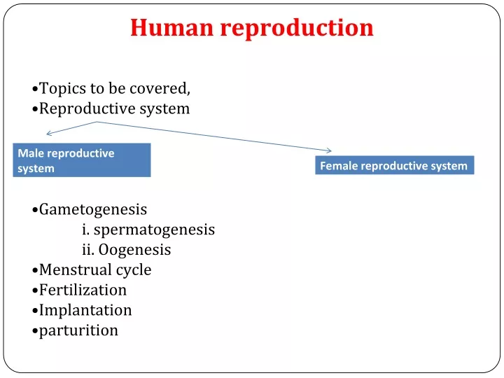
Human reproduction. Topics to be covered, Reproductive system Gametogenesis i. spermatogenesis ii. Oogenesis Menstrual cycle Fertilization Implantation parturition. THE MALE REPRODUCTIVE SYSTEM. Located in the pelvis region. Male reproductive system includes A pair of testes.

E N D
Human reproduction • Topics to be covered, • Reproductive system • Gametogenesis i. spermatogenesis ii. Oogenesis • Menstrual cycle • Fertilization • Implantation • parturition
THE MALE REPRODUCTIVESYSTEM. • Located in the pelvisregion. • Male reproductive systemincludes • A pair oftestes. • Accessoryducts. • Accessoryglands. • Externalgenitalia
Testes: • Located outside the abdominal cavity within a pouch called scrotum. • Scrotum provides low temperature required for spermatogenesis. • Each testis is about 4 to 5 cm length and 2 to 3 cm width. • Each testis has about 250 compartments called testicular lobules. • Each lobule contains one to three seminiferous tubules. • Seminiferous tubules lined by male germ cells and Sertoli cells. • Male germ cell undergoes meiosis and produce sperm. • Sertoli cells provide nutrition to the germ cell and the sperm. • In between the seminiferous tubule there is interstitial cell or Leydig cell. • Leydig cells produce testicular hormones • called androgen (testosterone).
Accessoryducts: • Includes rete testis, vasa efferentia, epididymis and vas deferens. • Seminiferous tubules open into vasa efferentia through rete • testis. • The vasa efferentia leaves the testis and open into epididymis. • The epididymis leads to vas deferens that ascends to the abdomen through inguinal canal and loops over the urinary bladder. • Vas deferens receives a duct from seminal vesicle and opens into the urethra as the ejaculatory duct. • Urethra originates from the urinary bladder and extends through the penis to its external opening called urethral meatus.
Accessoryglands: • Includes • Paired seminal vesicle • A prostate gland • Paired bulbourethral gland or Cowper’s gland • Secretion of these glands constitutes the seminal • plasma. • Seminal plasma rich in fructose, calcium, and certain enzyme. • Secretion of bulbo-urethral glands helps in • lubrication of penis.
Externalgenitalia: • Penis is the external genitalia. • It is made of special tissue that helps in erection of the penis to facilitate insemination. • The enlarged end of penis is called glans penis. • Glans penis is covered by a loose fold of skin • called foreskin.
THE FEMALE REPRODUCTIVESYSTEM • Located in the pelvic region of the female. • The female reproductive system includes: • A pair of ovaries • A pair of oviduct. • Uterus • Cervix • Vagina • External genitalia. • A pair of mammary • gland
Ovaries: • It is the primary female sex organs that produce the female gamete (ovum). • It also produces several steroid hormones. • The ovaries located in the lower abdomen. • Each ovary is about 2-4 cm in length. • Connected to the pelvic wall and uterus by ligaments. • Each ovary is covered by thin epithelium which encloses the ovarian stroma • The ovarian stroma has two zones • A peripheral cortex. • An inner medulla.
Oviduct: • Oviducts, uterus and vagina constitute the female accessory ducts. • Each fallopian tube is about 10-12 cm long and extends from the • periphery of each ovary to the uterus. • Close to the ovary the oviduct has a funnel shaped structure called infundibulum? • The edges of the infundibulum possess finger-like projections called fimbriae, which helps in collection of the ovum after ovulation. • The infundibulum leads to a wider part of the oviduct called ampulla. • The last part of the oviduct is called isthmus which joined to uterus.
Uterus: • It is single and is calledwomb. • It is inverted pearshaped. • Attached the pelvic wall byligaments. • The uterus opens into vagina through a narrowcervix. • The lumen of cervix is called cervicalcanal. • Cervical canal along with vagina form the birthcanal. • The wall of the uterus has three layers oftissues • Perimetrium: external thinmembranous. • Myometrium: middle thick layer of smoothmuscles • Endometrium: inner glandularlayer. • Endometrium undergoes cyclical changes during menstrualcycle. • Myometrium exhibits strong contraction during delivery of thebaby.
Externalgenitalia: • It includes following structure: • Mons Pubis: cushion of fatty covered • by skin and pubic hair. • Labia majora: fleshy folds of tissue which extends down from the mons pubis and surrounds the vaginal opening. • Labia minora: are paired folds of • tissue under the labia majora. • Hymen: the opening of vagina is often covered partially by a membrane called hymen. • Clitoris: a tiny finger-like structure lies at the upper junction of two labia minora above the urethral opening.
GAMETOGENESIS: (formation ofgametes) • Spermatogenesis: • Formation of sperm from the germ cell in the testes is spermatogenesis. • The process begins at puberty. • Spermatogonia present in the lining of seminiferous tubules undergo mitotic division to increase their number. • Each spermatogonium is diploid (2n) which contain 46 chromosomes. • Innermost layer of spermatogonial becomes larger called primary spermatocyte. • Primary spermatocyte undergoes meiosis-I to form two equal haploid (n) secondary • spermatocytes (n). • Each secondary spermatocyte undergoes meiosis-II to form two equal, haploid spermatids. • Each primary spermatocyte produces four spermatids. • Spermatids transformed into spermatozoa (sperms) by the process called spermiogenesis. • The sperm head embedded in the Sertoli cell. • Release of sperm from the seminiferous tubule is called spermiation.
Mammaryglands: • Mammary gland consists of glandular tissue and fat. • Glandular tissue of each breast divided into 15-20 mammary lobes. • Mammary lobes contain cluster of cells called alveoli. • The cells of alveoli secrete milk, stored in the lumen of alveoli. • The alveoli open into mammary tubules. • The tubules of each lobe join to form a mammary duct. • Several mammary ducts join to form a wider mammary ampulla. • Mammary ampulla connected • to lactiferous duct, through which milk is sucked out.
Hormonal control ofspermatogenesis: • This process is initiated at puberty due to secretion of gonadotrophins releasing hormone (GnRH) • GnRH secreted form hypothalamus and stimulate anterior • pituitary to secrete two gonadotrophins. • Luteinizing hormone (LH) and • Follicle stimulating Hormone (FSH) • LH acts on Leydig cells and stimulates synthesis of • androgens. • Androgen stimulates spermatogenesis. • FSH acts on Sertoli cells and stimulates spermatogenesis in other ways.
Structure ofsperm: • Ultrastructure of sperm consists of a head, neck, a middle piece and a tail. • Whole body of sperm surrounded by plasma membrane. • The sperm head contain an elongated haploid nucleus. • Above the nucleus a cap like structure present called acrosome. • The acrosome contains enzymes which help in fertilization of ovum. • The middle piece contains mitochondria, which provide energy for movement of tail that facilitate sperm motility. • Human male ejaculates 200-300 million sperms during coitus. • 60 percent must have normal shape and size and 40 percent of them must show • vigorous motility. • Sperm released from seminiferous tubules enters into accessory ducts. • On their way fluids from seminal vesicle and prostate gland added which • collectively called as Semen. • The function of male accessory ducts and glands are maintained by testicular hormone androgen.
Oogenesis: • Formation of a mature female gamete or ovum is calledoogenesis. • Oogenesis starts during embryonic stage, 25th week of the fetal age. • Germinal epithelium of ovary divided mitotically to produce millions of gamete mother cell or oogonia. • No oogonia formed or added after birth. • Oogonia enters into meiosis-I and proceeds uptodiakinesis of Prophase-I and get suspended, at this stage calledprimary Oocytes. • Each primary oocyte surrounded by layers of granulose cells and then called primary follicle. • At puberty only 60,000 to 80,000 primary oocytes are left in each ovary. • After puberty primary follicles get surrounded by more layers of granulosa cells and • a new theca to formsecondary follicles. • The secondary follicle transformed into tertiary follicle, characterized by a fluid filled cavity calledantrum. • The theca layers organized into an inner theca interna and outer theca externa. • During the growth of primary follicle into tertiary follicle during puberty, the primary oocyte restarts its first meiotic division and completes it within tertiary follicle resulting two unequal haploid cells. • Large haploid cell is called secondary oocyte. • A tiny cell called first polar body.
CONT…. • The secondary oocyte retains bulk of the nutrient rich cytoplasm of primary oocyte. • The tertiary follicle having secondary oocyte • further changes into Graafian follicle. • The secondary oocyte surrounded by a new membrane, zonapellucida. • The secondary oocyte undergoes second meiotic division continued upto metaphase- II and get suspended until entry of sperm. • At this stage Graafian follicle releases secondary oocyte from the ovary by the process called ovulation. • On entry of a sperm into the secondary oocytes stimulates it to complete meiosis-II and there is formation of a haploid ovum and a second polar body (n).
Zona pellucida: • Thick membrane • - exists for sometimes post fertilization • - outer protective layer Corona radiata: --2 layers of cell -- attached to zona pellucida -- supply vital protein to cell Vitalline membrane: -- plasma membrane of the egg cell -- control entry and exit of substances ooplasm: -- cytoplasm of egg cell -- supply nutrients to growing embryo
Germinal vesicle: -- nucleus of the egg cell -- contain genetic material Germinal spot: - Nucleolus of the cell
Menstrualcycle: • Reproductive cycle of female primates is called menstrual cycle. • The first menstruation begins at puberty is called Menarche. • Menstrual cycle repeated at an average interval of • 28/29 days. • One ovum is released in the middle of each menstrual cycle.
Events during Menstrual cycle • Menstruation or Mentrual (bleeding ) phase • Follicular phase or proliferative phase • Ovulatory phase • Luteal phase or secretary phase
Menstrualphase: • 1st phase of menstrual cycle. • Menstrual flow occurs. • Lasts for 3-5 days. • Breakdown of endometrial lining and blood vessel. • Mucus and blood comes out through vagina. • It occurs only when ovum released but no fertilization. • Lack of menstruation is the indication of pregnancy.
What causes the menstrual flow? • An egg is released from either of the ovaries every 28 days. • -an unfertilized egg is alive for 24 hours after ovulation. • -uterus prepares to receive the foetus • - endometrium thickness • -- no fertilization endometrium breaks bleeding happens
Factors affecting menstruation • Pregnancy • Poor health • Stress – delayed menstruation • Change in environmental conditions • Diet
Follicular phase Events that takes place in the follicular phase: • follicular growth • regenerative of endometrium • secretion of LH and FSH • ovulation
FOLLICULAR GROWTH Folicular growth occurs in ovary: • Primary follicles • Secondary follicles • Tertiary follicles • Graffian follicles
Conti…… -secretion of LH reaches a peak value during middle of menstrual cycle. --graffian follicles reptures --ovulation takes places. The corpus albicans (Latin for "white body") is an ovarian scar composed of connective tissue that forms after the corpus luteum degenerates, a process called luteolysis.
Follicularphase: • Menstrual phase followed by follicular phase. • Primary follicle becomes Graafian follicle. • Regeneration and proliferation of uterine endometrium. • LH and FSH level increases gradually in follicular • phase. • Level of estrogen increases as it is secreted from growing follicle. • It lasts for 5-13 days.
Ovulatoryphase: • FSH and LH attain peak level in this period (14th day). • This is called LH surge, which induces rupture of Graafian follicle and release of ovum from the ovary called ovulation.
Lutealphase: • Remaining part of Graafian follicle transformed into corpus luteum. • Coupusluteum produces large amount of progesterone. • Progesterone maintains the uterine endometrium, and prepares it for implantation. • Thickness of uterine endometrium increase in many folds, due to proliferation. • If there is fertilization, corpus luteum grows further and • pregnancy continued, menstrual cycle stopped. • In the absence of fertilization corpus luteum degenerates. • Disintegration of endometrium leading to menstruation. • Menstrual cycle ceases around 50 years of age, • called menopause.
FERTILIZATION ANDIMPLANTATION: • During copulation (coitus) semen is released by the penis into the vagina is called insemination. • The motile sperm swim rapidly, pass through cervix, uterus and finally reach the junction of isthmus and ampulla(ammpullary-isthmic junction). • The ovum released from the ovary also transported to ampullary isthmic junction where fertilization takes place. • Fertilization only takes place if both sperm and ovum reach ampullary – isthmic junction simultaneously. • The process of fusion of a sperm and ovum is called fertilization. • Acrosome of sperm secretes enzymes helps in • penetration into the ovum.
CONT… • Once a sperm comes contact with the zonapellucida of ovum and induces the changes in the membrane that blocks the entry of additional sperms. • That ensures monospermy and prevents polyspermy. • Only one sperm fertilize with one ovum. • Entry of sperm into the ovum induces the ovum to complete its second meiotic division of secondary oocyte. • Meiosis-II is also unequal cytokinesis resulting production of one large ovum (ootid) and one small second polar body. • Haploid nucleus of sperm fused with the haploid • nucleus of ovum to form a diploid zygote.
Sexdetermination: • Sex of a baby has been decided during fertilization and in the zygote. • Sex is determined by the sex-chromosomes present in • gametes. • Human female contain two XX chromosomes. • Human male contain XY chromosomes. • All the female gametes produced with only ‘X’ chromosome. • Sperms produced by male, 50% with ‘X’ and 50 % with ‘Y’ chromosome. • After fertilization zygote either carries XX or XY chromosomes. • Zygote with XX chromosomes develop into female and with • XY chromosome develops into male
Implantation -result of fertilization -diploid single celled zygote -mitotic division takes place repeatedly. -zygote divide mitotically to form Blastomeres – group of cells. --morula ----embryo with 8-16 blastomeres Blastocyst Trophoblast Inner cell
The zygote undergoes a series of mitotic cell divisions called cleavage. • The stages of development are: • Fertilized ovum (zygote) • 2-cell stage • 4-cell stage • 8-cell stage • Morula • Blastula • Gastrula
Embryonic development What happens once the implantation is done? -placenta formation takes place. -placenta is the structural and functional link between mother and foetus. • PLACENTA FORMATION: • Placenta is formed in 8-9 days. • Placenta is formed of 2 components • Fetal placenta • Maternal placenta
Fetal placenta • It is formed from trophoblast of blastocyst. • Umblical cord • Keeps the mother to connect with the foetus. • Maternal placenta • Formed from mother’s body. • Finger like projection on trophoblast is called chrionic villi • It combine with uterine tissue is called placenta.
Role of placenta Umbilical cord: Naval string Arise from foetus abdomen and connects to the placenta Nutrients come into foetus from mother’s blood Waste go out from foetus into mother’s blood. Exchange of oxygen and carbon di oxide. Placenta acts as an endocrine tissue and produces several harmones like human chorionic gonadotropin (hCG) Human lactogen (hPL) estrogen progestron In later phase of pergnancy, it secret a hormone called relaxin.
Cleavage: • Repeated mitotic division of the zygote without growth resulting a multicellular ball like embryo is calledcleavage. • Cleavage starts soon after fertilization. • Daughter cells produced during cleavage are called blastomeres. • The product of cleavage is called Morula, which is 8 to 16 celled. • The morula continues to divide and grow and transformed into blastocyst. • The blastomeres in blastocyst arranged into an outer layer • called trophoblast and an inner mass of cells attached to trophoblast • called inner cell mass. • Trophoblast cells attached to the endometrium helps development of placenta. • Inner cell mass gets differentiated into the embryo. • After attachment the uterine cells divide rapidly and cover the • blastocyst. • Blastocyst completely embedded in the uterine endometrium. This is called implantation.
Pregnancy and embryonicdevelopment: • After implantation, finger like projections appears on the trophoblast called chorionic villi. • Chorionic villi surrounded by uterine tissue and maternal blood. • Temporary association between the fetal tissue (chorionic villi) and maternal tissue (uterine endometrium) is called placenta.
Function ofplacenta: • The embryo connected to the placenta by umbilical cord, which transports substances to and from the embryo. • Facilitate transport of oxygen and nutrient from mother to embryo. • Removes CO2 and waste material from the embryo. • Acts as endocrine gland and produces several hormones like: • Human chorionic gonadotrophins (hCG) • Human placental lactogen (hPL) • Estrogen. • Progesterone • Relaxin produced from the ovary in the later stage of pregnancy.