Download
1 / 47
480 likes | 785 Views
Human Reproduction. “If you study the divorces, as we have had to do in these past years you will find…sex is the first (reason). They did not get along sexually. They may not say that in court. They may not even tell that to their attorneys but that is the reason. Spencer W. Kimball,

E N D
“If you study the divorces, as we have had to do in these past years you will find…sex is the first (reason). They did not get along sexually. They may not say that in court. They may not even tell that to their attorneys but that is the reason. Spencer W. Kimball, (The teachings of Spencer W. Kimball, Salt lake city: Bookcraft, 1982, pg 46)
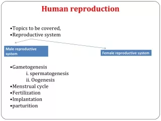
Sex Organs • Primary sex organs: • Are the gonads i.e.… Testes and Ovaries • Produce the gametes and sex hormones. • Accessory sex organs • Ducts that provide transport for gametes • Glands that provide nutrients
Male Reproductive System Testes • Formed in abdominal cavity. • Cryptorchidism “hidden testes” • Sterility if not corrected in childhood • Subject ot tumor formation
SCROTUM Outpouching of skin that contains both testes; can be moved closer to or farther from body to help maintain temperature suitable for sperm formation
Testes • Cutting inside the testes you would find 125 meters of inter-connecting tubes called • Seminiferous tubules
Seminiferous tubules • The location in which germ cells undergo meiosis to become sperm.
Spermatogenesis • Sperm are derived from 2n spermatogonium germ cells. • As these cells divide they are pushed away from the wall into the tubules interior. • Spermatogenesis takes about 9-10 weeks • Spermatogenesis is controlled by sertoli cells. • Seritoli cells also provide nourishment for the developing sperm cells. Sertoli cell Spermatogonium Germ cell
Sperm • The sperm cell can be divided into three parts: tail, midpiece and head. • The tail is a long singular falgella • Midpiece moves the flagella and has many mitochondria • Head contains DNA • Acrosome contains digestive enzymes which are release to unable the sperm to work its way through the zona palucida. acrosome Head Tail Midpiece
Testosterone • Are produce by cells inside the testes called Leydig cells • Functions of Testosterone • Development of sperm • Development of secondary sex characteristics • Traits that are distinctly associated with maleness but do not play a direct role in reproduction example: voice, shoulder, muscle, body hair.
Epdidymis • A duct located on top of the testis • Functions as a place in which sperm are stored and where they mature. Epididymis
Vas Deferens • One of a pair of ducts for rapid transport of sperm • Vasectomy is a common surgical method of sterilizing males. • Males ejaculate without semen • Semen is the glandular secretion which become mixed with sperm Vas Deferens
Seminal Vesicle • One of a pair of glands that secrete fructose sugar as part of the semen • Makes the semen very viscous Seminal vesicle
Prostate gland • Contributes prostate fluid, to the semen. • Prostate fluid is highly alkaline to off-set the high acidity of the vagina. Prostate gland
Bulbourethral glands Upon sexual excitement and prior to ejaculation, these glands are stimulated to secrete a mucoid substance which coats the lining of the urethra to neutralize the pH of the urine residue and lubricates the tip of the penis in preparation for sexual intercourse Bulbourethral (Cowper’s) gland
Urethra • Serves as a common tube for both the urinary and reproductive systems. Urethra
Penis • Composed of erectile tissue which contains numerous vascular spaces that become engorged with blood. • Sexual arousal leads to an erection in which the penis becomes longer, wider and firmer. • An erection is absolutely necessary in order to have normal sexual intercourse. Penis
Erectile Dysfunction • Impotence is the inability of a man to have an errection. • Causes may include • Injury to nerves, arteries, are muscles • Medications for high blood pressure, antihistamines, and depression • Psychological factors such as anxiety, guilt, depression, and fear of sexual failure cause up to 20 % of all cases
Hormonal control of male reproduction Fig. 45.5, p. 787
Primary Sex Organ The Ovaries
Ovaries • Each ovary produces an ovum about every other month • The ovaries stagger their production so that one ovary produces an ovum one month and the other produces it the following month.
Menstrual Cycle • The menstual cycle is a recurring reproductive cycle, which last on average 28 days. Durring this time an oocyte is released from one of a pair of ovaries, and the endometrial lining of uterus is primed for pregnancy. • In the event that the oocyte does not become feritilized endomytrial tissue along with blood from the uterus will flow out through the vaginal canal (menstruation).
secondary oocyte Ovulation. Mature follicle ruptures and releases the secondary oocyte and the first polar body. first polar body antrum A corpus luteum forms from remnants of the ruptured follicle. A primordial follicle; meiosis I has been arrested in the primary oocyte inside it. When no pregnancy occurs, the corpus luteum degenerates. primordial follicle Fig. 45.7b, p. 790
Follicle Production Ovulation Mature follicle • At birth, a female will have all the oocytes she will ever have. • At puberty a single oocyte will mature inside a follicle. • The follicle consist of granulosa cells that produce estrogen and an oocyte. • From day 1 through day 13 the follicle grows producing more and more estrogen. • Estrogen causes the production of Follicle stimulating hormone (FSH). • FSH cause the follicle to grow which in turn produces more estrogen (positive feedback) Granulosa cells A primordial follicle; meiosis I has been arrested in the primary oocyte inside it.
Ovulation • In Large amounts, estrogen also stimulates the production of Luteinizing hormone (LH). • LH causes the follicle to rupture and the oocyte to burst out. This process is called “ovulation” • In a 28 day menstrual cycle ovulation occurs on day 14. • It is at this time that a woman is most likely to become pregnant.
Development of the Corpus Luteum • After ovulation, the granulosa cells in the ruptured follicle come together to form a structure called the corpus luteum. • The corpus luteum’s function is to secretes progesterone. • Progesterone keeps the uterine lining in place for implantaiton of the egg.
Fertilization • The oocyte is released and is taken up by the oviduct. • The cells lining the oviduct are ciliated and move the oocyte toward the uterus • Successful Fertilization of the oocyte occurs in the upper portion of the oviduct
Fertilization cont. • Out of about 200 million sperm only about 50 – 100 reach the egg. • When sperm contact the oocyte they release enzymes that clear a path through the zona pellucida. • As soon as the first sperm penitrates the plasma membrane the zona pellucida instantly hardens keeping all other sperm out. • The fusion of the sperm and egg create a zygote. The zygote is 2n. Zona pellucida
Oviducts • As the zygote travels down the oviduct it undergoes cleavage. • Cleavage is the multiplication and division of cells in the zygote without the zygote growing in size. • The zygote stays the same size as the cells divide into smaller and smaller units • By the fifth day the zygote is a ball of cells called a blastocyst. • After 7-10 days old the blastocyst implants itself in the uterus.
The Uterus • consists of 2 major layers: the myometrium and the Endometrium. • Myometrium is a large muscle layer that is able to contract for long periods of time. • Endometruim is the inner mucosal lining that thickens and sloughs off a periodic cycles during the menstral cycle. It is also the layer where the blastocyst implants. Myometrium Endometrium Cervix
Blastocyst • The Blastocyst consist of two different cell types that form in inner and outer layer. • The inner layer of cells produce the embryo. • The outer layer of cells called the trophoblast implant into the uterine wall and produce the placenta. Trophoblast Endometrium Inner cell mass
Ectopic pregnancy • If the blastocyst implants somewhere outside the uterus it is called an ectopic pregnancy. • Implantation in the uterine tube is called a tubule pregnancy. This results in a medical emergency. • Implantation can also occur in the intestines or the walls lining the abdominopelvic cavity
MATERNAL CIRCULATION FETAL CIRCULATION embryonic blood vessels maternal blood vessels movement of solutes to and from maternal blood vessels (arrows) umbilical cord blood-filled space between villi chorionic villus tissues of uterus fused amniotic and chorionic membranes Fig. 45.13a, p. 797
4 weeks 8 weeks 12 weeks appearance of the placenta at full term Fig. 45.13b, p. 797
OVIDUCT OVARY UTERUS MYOMETRIUM ENDOMETRIUM urinary bladder opening of cervix urethra CLITORIS LABIUM MINOR LABIUM MAJOR anus VAGINA Fig. 45.6b, p. 789
Vagina • Tube that receives sperm. • The lower part of birth canal. • Muscular with folds so that is can extend. • Sexual arousal stimulates mucus secreting glands that lubricate the vagina in preparation for sexual intercourse. Vagina
Vulva • Consists of • Labia major • Labia minor • Clitoris Clitoris Labia minor Labia major
secondary oocyte surrounded by follicle cells surface of ovary Fig. 45.8, p. 791
hypothalamus GnRH anterioir pituitary FSH LH midcycle peak of LH (triggers ovulation) hypothalamus Blood levels of FSH (purple) and LH (lavender) anterior lobe of pituitary gland FSH LH LH growth of follicle ovulation corpus luteum estrogens progesterone, estrogen Blood levels of estrogens (light blue) and progesterone (dark blue) estrogens progesterone, estrogen endometrium of uterus menstruation Fig. 45.9, p. 792 Days of one menstrual cycle (using 28 days as the average duration) FOLLICULAR PHASE OF MENSTRUAL CYCLE LUTEAL PHASE OF MENSTRUAL CYCLE