Download
1 / 39
390 likes | 711 Views
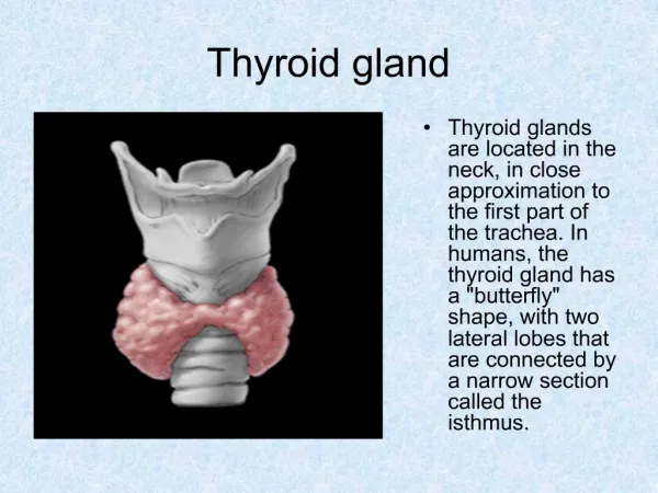
In the name of God. Digestive gland. Dr. Zahiri. Digestive Glands. Digestive glands are consist of: Salivary glands Pancreas Liver Gall bladder. Salivary Glands. minor (Accessory ) salivary glands pairs of major (main) salivary glands Three pairs of major salivary glands are:

E N D
In the name of God Digestive gland Dr. Zahiri
Digestive Glands Dr. Maria Zahiri Digestive glands are consist of: • Salivary glands • Pancreas • Liver • Gall bladder
Dr. Maria Zahiri Salivary Glands • minor (Accessory) salivary glands • pairs of major (main) salivary glands • Three pairs of major salivary glands are: • Submandibular glands • Sublingual glands • Parotid glands
Salivary Glands Dr. Maria Zahiri • capsule and septa • that organize the glands into lobes and lobules
Salivary Gland Cells Dr. Maria Zahiri • Serous cells • produce proteins • pyramidal cells • spherical euchromatic nucleus • Cytoplasm is basophilic (RER, Golgi apparatus) • Apically located granules that are usually eosinophilic • Many basal mitochondria
Dr. Maria Zahiri • Mucous cells • cuboidal or pyramidal • heterochromatic basal flattened nucleus • Apical cytoplasm is pale • Theyhave less RER, fewer mitochondria but greater GA
Dr. Maria Zahiri • Organized as acinus or demilune that secrete proteins, polysaccharides and ptyalin
Dr. Maria Zahiri • Ducts system • Intercalated duct • smallest branches • short, cuboidal or squamous cells and myoepithelial cells • Striated duct • Largerdiameter • cuboidal to low columnar cells which are eosinophilic, and secrete fluid and ions • basolateral membrane folded with Na – ATPase pump, elongated mitochondria
Dr. Maria Zahiri • intralobularducts: • Striated ducts join each other and forming intralobular ductsthat are invested by more CT elements • interlobular ducts (excretory ducts): • intralobular ductsjoin each other and forming larger caliber ducts known as interlobular ducts (excretory ducts) • Excretory duct have large diameter, large lumen, cuboidal or columnar cells, located outside of lobules
Dr. Maria Zahiri • Myoepithelialcells (Basket cells) • have large processes which form desmosomal contact with acini and ducts cells, • Their processes are rich in actin and myosin • have a common basal lamina with acinar cells • They attach to basal lamina by hemidesmosome
Parotid Gland Dr. Maria Zahiri • The largest salivary gland • produce 30% of saliva • capsule & many septa divided the glands into lobes and lobules • serous cells • After 40 year of age adipose • tissue invaded the gland
Submandibular Gland Dr. Maria Zahiri • produce 60% of saliva • Mucous and serous acini; mucous acini with limited number of serous demilunes • About 80% of cells are serous (basophilic)
Sublingual Gland Dr. Maria Zahiri • It is very small that produce 5% of total saliva • Mostly mucous cells in acini with some serous demilunes • Produce mix saliva but mostly mucous saliva
Saliva Dr. Maria Zahiri • Saliva includes water, enzymes, IgA, mucous, ions • Moisten and lubricate food for swallowing • Enzymes like amylase and lipase to begin digestion • Saliva has protective effect on oral cavity tissues • Participate in taste sensation • IgA, lactoferrin, lysozyme have different role against antigens and microorganisms
Pancreas Dr. Maria Zahiri • is a mixed exocrine and endocrine gland • Thin capsule with septa between lobules • Exocrine part organized similar to parotid gland • Endocrine part are islets of Langerhans scattered among the excretory units • Centroacinar cells occupy the lumen of the acini, these cells are beginning of the duct system • Centroacinar cells are pale, low cuboidal • Intercalated ducts, intercalated ducts, Intralobular ducts, and interlobular ducts
Liver Dr. Maria Zahiri • largest gland in the body (1500 gr) • has endocrine and exocrine functions • Receives portal blood from intestine via portal vein and oxygenated blood from hepatic arteries
Dr. Maria Zahiri Liver • hepatocytes • Liver has a lobular organization • Classical lobule: hepatocytes arranged as an hexagon • Portal area (triad): • is where three classical lobules are in contact with each other, more CT elements present, • houses branches of hepatic artery, • tributaries of portal vein, • interlobular bile ducts, and lymph • vessels
classical lobule • blood flow from periphery to center of lobule • Bile flow in opposite direction in small intercellular spaces known as bile canaliculi • Portal lobule • Is a triangular region that portal area is located in its center and central veins form apices of the triangle • Hepatic acinus (acinus of Rappaport) • is diamond-shaped, a distributing artery located in center of acinus, three regions of parenchyma surrounding the artery(zone I, II, III)
Liver • The space between the anastomosing plates of hepatocytes are occupied by hepatic sinusoids • Sinosuidal lining cells are fenestrated are not in contact with each other They prevent direct contact between blood and hepatocytes • Resident macrophages known as Kupffer cells associated with lining cells • The space that separates sinusoidal lining cells from hepatocytes is called perisinusoidal space (space of Disse) • Microvilliof hepatocytes occupy much of the space • Type III collagen that is present in space support lining cells(no basal lamina)
Cells • Fat storing cells ( known as Ito cells or Stellate cells) may present in Disse space and store vitamin A • Pit cells which are natural killer cells also may be seen in space of Disse • Hepatocytes are polygonal • forming anastomosing plates of one to two thickness cells, eosinophilicwith 1 or 2 spherical nuclei,
Cells • In lateral domain bile canaliculi form between two cells, microvilli, Na-K ATPase and gap junctions are common characteristics of lateral domain • Bile canaliculi between hepatocytes leading to hepatic ducts with simple cuboidal epithelium
Liver • Sinunosoidal domains of cell membrane have many microvilli protrude to perisinusoidal space (of Disse), endocrine secretion of hepatocytes release here • Bile secreted from hepatocytes into bile canaliculi contains water, ions, bile salts and acids, phospholipids, cholesterol, & bilirubin
Liver Functions • Glycogen storage • Lipid metabolism • Vitamin storage (A, D, B12 ) • Bile production (Bile acids solubilize lipids and aid digestion) • Detoxification of drugs and toxins in SER • Synthesis of plasma proteins (albumin) • Metabolism of lipid, carbohydrate, proteins • Erythrocyte breakdown (Bilirubin formed in breakdown of RBC) • Complex IgA with secretory component
Gall Bladder • Mucosa : simple columnar epithelium • Epithelial layer is highly folded • lamina propria: • loose CT • Smooth muscle layer is composed of thin obliquely oriented fibers with perimuscular connective tissue • Serosal and adventitial membrane • Stores and concentrates 30-50 ml bile • Cholecystokinin and acetylcholine stimulates contraction to force bile into small intestine
Dr. Maria Zahiri با آرزوی روزگاری خوش