Download
1 / 52
630 likes | 1.31k Views
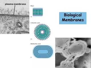
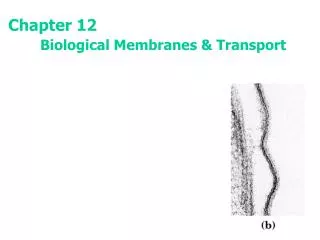
Biological Membranes. Chapter 11 (Page 369-383). Electron Micrograph of Biological Membranes. 1. Biological Membranes. In eukaryotic cells, membranes play many important functions. Define the external boundaries of cells and regulate the molecular traffic across that boundary.

E N D
Biological Membranes Chapter 11 (Page 369-383)
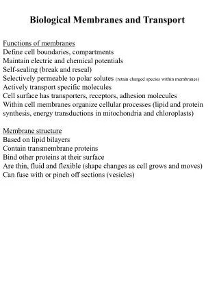
1. Biological Membranes • In eukaryotic cells, membranes play many important functions. • Define the external boundaries of cells and regulate the molecular traffic across that boundary. • Divide the internal space into discrete compartments to segregate processes and components. • Aid in cell-to-cell communication and in signaling. • Organize complex reaction sequences and cellular processes. • Energy transduction • Biomolecule synthesis
2. General Physical Properties of Membranes that enable their Biological Activities • Very thin (3 to 10 nm) • Flexible • Self-sealing • Selectively permeable to polar solutes
Lipid-Water Interactions lead to Cell Formation
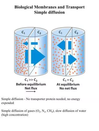
1. Lipids Aggregate into Structures in Water • Lipids are typically amphiphatic molecules that are water insoluble. When mixed with water they: • Aggregate in a phase separate from their aqueous surroundings. • The hydrophobic (nonpolar) moieties cluster together to reduce exposure to water. • The hydrophilic (polar) groups interact with the surrounding water. • Greater entropy for these water molecules than at the water-nonpolar moiety interphase.
1. Lipids Aggregate into Structures in Water • Thermodynamically driven by hydrophobic interactions, three types of structure can form that depend on: • Type of lipid • Concentration • Micelle • Bilayer • Liposome
2. Micelle Formation Hydrophilic exterior interacts with water Hydrophobic interior; water excluded
2. Micelle Formation • A micelle forms in the solution of amphipathic molecules that have larger polar head than nonpolar tail • Fatty acids • Sodium dodecyl sulfate • Aggregation occurs when the concentration of molecules is higher than a certain threshold • Each micelle has from a few dozen to a few thousand lipid molecules
3. Bilayer Formation Hydrophilic exterior interacts with water Hydrophobic side transiently interacts with water Hydrophobic interior; water excluded
3. Bilayer Formation • A bilayer forms when the cross-sectional areas of the head group and nonpolar tail are similar • Glycerophospholipids • Sphingolipids • Because the hydrophobic regions at the edges are transiently in contact with water, the bilayer sheet is unstable
4. Liposome Formation • A liposome forms by a bilayer spontaneously folding back on itself to form a hollow sphere (a vesicle) • By forming liposomes, bilayers lose their hydrophobic edge regions and achieve maximal stability in their aqueous environment. • A separate aqueous compartment is formed. • Liposomes can be seen as precursors to the 1st living cells.
The Composition of Eukaryotic Biological Membranes
1. What are Membranes? • Complex lipid-based pliable structures composed of a variety of lipids and proteins. • Some membrane lipids and proteins are glycosylated. • All cells have a cell membrane, which separates the cell from its surrounding. • Eukaryotic cells have various internal membranes (organelles) that divide the internal space into compartments. • - NOTE: Mammalian red blood cells (erythrocytes) do not have organelles. • Why???? • Perhaps to make room for hemoglobin.
2. Common Features of Eukaryotic Membranes A. The membrane of eukaryotic cells consists of two leaflets of lipid-based monolayers: • One leaflet faces the cytoplasm • One leaflet faces the extracellular space or the inside of membrane-enclosed organelle
2. Common Features of Eukaryotic Membranes • B. Sheet-like flexible structure, 3–10 nm thick • C. Structures within the membrane bilayer are stabilized by noncovalent forces, especially hydrophobic ones • D. Membrane bilayers are largely composed of phospholipids. • The polar heads are on the exterior forming a hydrophilic surface. • The fatty acyl chains are in the interior forming a fluid, hydrophobic region. • Other lipids are nestled in between.
2. Common Features of Eukaryotic Membranes • E. Protein molecules span the lipid bilayer • F. Asymmetry • Some lipids are found preferably “inside” • Some lipids are found preferably “outside” • Carbohydrate moieties are always outside the cell • Electrically polarized (inside negative ~ –60mV) • G. Impermeable to polar solutes • Specific transporters allow transport
3. Fluid Mosaic Model of Membranes • The combination of electron microscopy and chemical composition studies, and physical studies of permeability and the motion of individual protein and lipid molecules within the membranes led to the development of the fluid mosaic model (Singer and Nicholson, 1972). • Lipids form a viscous, two-dimensional solvent into which proteins are inserted asymmetrically. • -Membrane sidedness • Integral proteins are firmly associated with the membrane via nonpolar sidegroups, often spanning the bilayer. • - Some have multiple transmembrane domains • Peripheral proteins are weakly associated (noncovalently) and can be removed.
3. Fluid Mosaic Model of Membranes Membrane mosaic is fluid because the noncovalent interactions enable the molecules to freely move laterally.
4. Composition of Membranes • The composition of membranes is different in: • different organisms, tissues, and organelles • Ratio of lipid to protein varies • Type of lipid varies • Phospholipid and sterol types vary • Galactolipids abundant in plant chloroplasts but almost absent in animals • Type of protein varies • Some membranes have a predominance of only one protein; specialized function
4. Composition of Membranes More than 90% of the rod cell plasma membrane of the retina is made of the light-absorbing glycoprotein rhodopsin. Webvision.umh.es/websvision/sretina.html
4A. Membrane Composition is Highly Variable in Different Organisms
4B. Membrane Composition is Highly Variable in Different Organelles • Phospholipids are abundant in all membranes. • Cholesterol is abundant in the plasma membrane.
5. Membrane bilayers are Asymmetric. Every component of the membrane exhibits asymmetry • Lipids • Outer and inner leaflets have different lipid compositions • Proteins • Individual peripheral membrane proteins are only associated with one side of the membrane • Integral membrane proteins have different domains on different sides of the membrane. • Specific lipid modification of proteins targets the protein to a specific leaflet • Carbohydrates • Only on the outside of cells
5A. Membrane bilayers are Asymmetric in Lipid Composition • The two leaflets of membranes have different lipid compositions: • Positioning of a lipid on either leaflet can serve a functional role • Phosphatidylserine (typically positioned in the inner leaflet) on the outer leaflet of the plasma membrane serves to • Activate blood clotting (platelets) • Mark the cell for destruction (other cells)
5AI. Asymmetry in Erythrocytes • Choline-containing lipids are typically found in the extracellular leaflet.
6. Two Main Types of Membrane Proteins Peripheral proteins Integral proteins
6A. Peripheral Membrane Proteins • Linkages with the membrane: • Associate with the polar head groups of membranes • Relatively loosely associated with membrane • Through ionic interactions with the lipids or aqueous domains of integral membrane proteins B. Removed by disrupting ionic interactions or hydrogen bonds either with high salt or change in pH.
6A. Peripheral Membrane Proteins C. May serve as regulators of membrane-bound enzymes. D. May limit mobility of integral proteins by tethering them to intracellular structures. E. Purified peripheral membrane proteins are no longer associated with any lipids.
6B. Integral Membrane Proteins A. Span the entire membrane B. Localized asymmetrically • Different domains in different compartments • Molecules of an ion pump have the same orientation and therefore pump in the same direction.
6B. Integral Membrane Proteins C. Tightly associated with membrane • Hydrophobic stretches in the protein interact with the hydrophobic regions of the membrane D. Removed by detergents that disrupt the membrane E. Purified integral membrane proteins still have phospholipids associated with them
6C. Function of Integral Membrane Proteins A. Receptors: Detecting signals from outside • Light (opsin) • Hormones (insulin receptor) • Neurotransmitters (acetylcholine receptor) • Pheromones (taste and smell receptors) B. Channels, gates, pumps for transport • Nutrients (maltoporin) • Ions (K-channel) • Neurotransmitters (serotonin reuptake protein) C. Enzymes • Lipid biosynthesis (some acyltransferases) • ATP synthesis (F0F1 ATPase/ATP synthase)
6D. Six Types of Integral Membrane Proteins Type I and II: Have only one transmembrane helix; the amino- terminal domain is outside the cell in type I and inside in type II. Type III: Have multiple transmembrane helices in a single polypeptide. Type IV: Transmembrane domains of several different polypeptides assemble to form a channel through the membrane. Type V: Proteins are held to the bilayer primarily by covalently linked lipids. Type VI: Proteins have both transmembrane helices and lipid (GPI) anchors.
6E. Determining Membrane Protein Topology Membrane protein topology refers to determining the 3-D structure of a membrane protein and also the location of domains with respect to the lipid bilayer. • X-ray crystallography • Once a daunting task for membrane proteins is becoming more commonplace with sophisticated new approaches. B. Reactions with reagents to identify extracellular domains • Membrane-impermeant reagents such as trypsin. C. Hydropathy index measurements to identify transmembrane domains. These approaches require sequencing these proteins using standard tecniques.
6EI. Amino Acids in Membrane Proteins Cluster in Distinct Regions • Transmembrane segments are predominantly hydrophobic • Tyr and Trp cluster at nonpolar/polar interface • Charged amino acids are only found in aqueous domains
6EII. Structural Motifs of Transmembrane Segments Helices β Barrels • An helical sequence that spans the membrane contains 20 to 25 residues. • 7 to 9 residues of the β conformation span the membrane. How are these motifs different than their corresponding structures in soluble proteins?
6EII. Structural Motifs of Transmembrane Segments • In soluble proteins, the side groups of the amino acids that participate in helices and β barrels tend to be polar. • - Interact via hydrogen bonds with water. • In membrane proteins, there is a higher nonpolar content in the side groups of transmembrane domains. • - In the absence of water the domains form helices and β barrels to maximize intrachain H-bonds.
6EII. Structural Motifs of Transmembrane Segments • The side groups in helices are largely nonpolar. • Due to alternating side chains projecting above and below βsheets, every second residue in β barrels is hydrophobic and interacts with the lipid bilayer and the other residues may or may not be hydrophilic.
6EIII. The Hydropathy Index can Predict the Topology of Membrane Proteins • The hydropathy index is a calculation used to determine sequences of hydrophobicity and hydrophilicity in membrane proteins. • The hydropathy index (Table 3.1) is a measure of the relative polarity of each amino acid. • Positive values: Hydrophobic • Negative values: Hydrophilic • The hydropathy index for a sequence is calculated by averaging the hydropathy index for all amino acids within a set window of amino acids.
6EIII. The Hydropathy Index can Predict the Topology of Membrane Proteins • A window size of 5-7 amino acids is good for finding hydrophilic regions that are likely exposed on the surface. • A window size of 19-21 AA is good for finding hydrophobic, membrane-spanning domains. • This method works well for -helical transmembranes but not for β barrels. • Let’s focus on applying this method for finding hydrophobic domains.
6EIIIa. HydropathyPlot Determination Kyle-Doolittle Scale
6EIIIa. HydropathyPlot Determination • For a given window size, say 20 AA and a protein of 160 AA residues: • Calculate HI(1-20) by averaging the HI for AA1 through AA20 then plot for middle residue (AA10). • Calculate HI(2-21) by averaging the HI for AA1 through AA20 then plot for middle residue (AA11). • Continue until you reach sequence 141-160 and plot for residue AA150. • Any region that exceeds +1.6 is likely a transmembrane segment.
Hydropathy Plot Determination for Transferrin Receptor 1 (TfR1) dimer axes Type II Plasma Membrane Ectodomain (AA 89 – 760)) Transmembraneregion (AA68 - 88) Cytoplasmic region (AA1 - 67)
Hydropathy Plot Determination for Transferrin Receptor 1 (TfR1) • Obtain protein sequence from http://www.uniprot.org website. • Search in: Protein Knowledge (UnitProtKB) • Query: Transferrin Receptor 1 • Click on Homo Sapiens Entry (Q9UP52) • Sequence: Copy the sequence • 1 MMDQARSAFSNLFGGEPLSYTRFSLARQVDGDNSHVEMKLAVDEEENADNNTKANVTKPK • 61 RCSGSICYGTIAVIVFFLIGFMIGYLGYCKGVEPKTECERLAGTESPVREEPGEDFPAAR • 121 RLYWDDLKRKLSEKLDSTDFTGTIKLLNENSYVPREAGSQKDENLALYVENQFREFKLSK • 181 VWRDQHFVKIQVKDSAQNSVIIVDKNGRLVYLVENPGGYVAYSKAATVTGKLVHANFGTK • 241 KDFEDLYTPVNGSIVIVRAGKITFAEKVANAESLNAIGVLIYMDQTKFPIVNAELSFFGH • 301 AHLGTGDPYTPGFPSFNHTQFPPSRSSGLPNIPVQTISRAAAEKLFGNMEGDCPSDWKTD • 361 STCRMVTSESKNVKLTVSNVLKEIKILNIFGVIKGFVEPDHYVVVGAQRDAWGPGAAKSG • 421 VGTALLLKLAQMFSDMVLKDGFQPSRSIIFASWSAGDFGSVGATEWLEGYLSSLHLKAFT • 481 YINLDKAVLGTSNFKVSASPLLYTLIEKTMQNVKHPVTGQFLYQDSNWASKVEKLTLDNA • 541 AFPFLAYSGIPAVSFCFCEDTDYPYLGTTMDTYKELIERIPELNKVARAAAEVAGQFVIK • 601 LTHDVELNLDYERYNSQLLSFVRDLNQYRADIKEMGLSLQWLYSARGDFFRATSRLTTDF • 661 GNAEKTDRFVMKKLNDRVMRVEYHFLSPYVSPKESPFRHVFWGSGSHTLPALLENLKLRK • 721 QNNGAFNETLFRNQLALATWTIQGAANALSGDVWDIDNEF
Hydropathy Plot Determination for Transferrin Receptor 1 (TfR1) • Go to http://www.vivo.colostate.edu/molkit/hydropathy/ • Insert the sequence into the window • Specify window size • Specify Kitt-Doolittle scale One transmembrane region correctly predicted at the N- terminus
Hydropathy Plot Correctly Predicts 7 Transmembranes for Bacteriorhodopsin
6F. Lipid Anchors • Some membrane proteins are lipoproteins. • They contain a covalently linked lipid molecule • Long-chain fatty acids • Isoprenoids • Sterols • Glycosylated phosphatidylinositol (GPI) • The lipid part can become part of the membrane • The anchoring process is reversible. • May be more than one attached lipid moiety • Other interactions, such as ionic attractions between positively charged Lys residues in the protein and negatively charged head groups contribute stability.