Download
1 / 43
550 likes | 1.25k Views
Renal Tubular Acidosis. Jeffrey J Kaufhold MD FACP February 2010. Summary. Review of renal tubule physiology Types of RTA’s Why they are named the way they are Subtypes Comparison and constrasting of RTA’s Treatment. Glomerular Physiology. Filtration Filtration membrane

E N D
Renal Tubular Acidosis Jeffrey J Kaufhold MD FACP February 2010
Summary • Review of renal tubule physiology • Types of RTA’s • Why they are named the way they are • Subtypes • Comparison and constrasting of RTA’s • Treatment
Glomerular Physiology • Filtration • Filtration membrane • Endothlial cell layer • Basement membrane • Epithelial cell layer • Electrical charge – negative • Clearance = waste product removal • Ultrafiltration = water removal
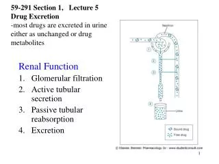
Renal Tubule PhysiologyOverview Prox. tubule Distal Tubule reabsorption Collecting duct Loop of Henle
Proximal Tubule • Function: Reabsorption • Features: • Brush border with cilia • Carbonic Anhydrase for reclaiming Bicarb • Filtration Fraction • Pathology: Renal tubular Acidosis
Renal Tubule PhysiologyOverview Carbonic Anhydrase Prox. tubule Distal Tubule blood Tubule membrane reabsorption Collecting duct Ultrafiltrate Tubule lumen Loop of Henle Tubule membrane
Renal Tubule PhysiologyOverview Prox. tubule Distal Tubule Collecting duct Loop of Henle
Renal Tubule PhysiologyOverview Prox. tubule Distal Tubule Collecting duct impermeable to imperm. to H2O solute Loop of Henle
Renal Tubule PhysiologyOverview Prox. tubule Distal Tubule Collecting duct Loop of Henle
Renal Tubule PhysiologyOverview Ion Exchange Sodium for Potassium/Hydro Prox. tubule Distal Tubule Collecting duct Loop of Henle
Renal Tubule PhysiologyOverview K+ H+ Ion Exchange Sodium for Potassium/Hydro Prox. tubule Distal Tubule Na+ Loop of Henle
Renal Tubule PhysiologyOverview Prox. tubule Distal Tubule solute exchange reabsorption Collecting duct impermeable to imperm. to H2O solute ADH + permeable to H2O Loop of Henle ADH - impermeable
Interstitium • The tissue in between the tubules • Function: • Ammoniagenesis • Dependent on renin, aldosterone. • NH3 converted to NH4Cl provides a huge sink for nontitratable acid Disorders of ammoniagenesis occur in severe hypOkalemia and Diabetic Nephropathy.
RTA’s • Named according to order in which they were described, i.e. severity. • Type I is worst showing up in children with failure to thrive, Ricketts, hypokalemia, stones • Type II also shows up in children, but not as severe. • Type III rare • Type IV not described until diabetics lived long enough to develop renal comps.
Renal Tubular Acidosis • Type I most severe, occurs in the Distal tubule, and is congenital problem with the transport proteins responsible for excretion of acid. • Four types of Type I distal RTA: • Rate dependent (defective or decreased pump) • Secretory (absent proton pump), • Gradient dependent (Backleak of K) • Voltage dependent (defective K channels) • Seen in sickle cell, obstructive uropathy • Potassium channels affected by Amiloride, lithium • Bicarb can go as low as 8 • HypOkalemia (hypERkalemia in voltage dependent)
Renal Tubular Acidosis • Type II Proximal Tubular Acidosis • Less severe, immaturity of proximal tubule leads to bicarb loss. • Corrects during puberty • Bicarb usually 15 or greater • HypOkalemia • Large bicarb requirement • Stones, Failure to thrive. • 11 types associated with conditions such as myeloma, Fanconi’s syndrome.
Type II Proximal RTA subtypes • Hereditary • Fanconi syndrome • Wilson’s dz, Cystinosis • Tyrosinemia, PyruvateCarboxylase Deficiency • Acquired • 1. Drugs (TCN, Gent, Glue sniffer, GMP) • 2. Heavy Metals • 3. Immunologic disease (sjogrens, Myeloma) • 4. Balkan nephropathy • 5. Nephrotic syndrome/ transplant dysfunction • 6. Osteoporosis • 7. PNH
Renal Tubular Acidosis • Type III • Small kindred of children born with Combo of type I distal RTA AND tubular immaturity of proximal tubule type II RTA. Have features of both, and the proximal tubule disorder corrects after puberty, leaving them with a true Type I distal RTA.
Renal Tubular Acidosis • Type IV RTA • Also known as Hypoaldosteronism, Hyporeninism. • Problem with ammoniagenesis • Commonly seen in Diabetics, Sarcoidosis, Chronic Pyelonephritis, Gouty nephropathy, chronic rejection • Bicarb as low as 18, may be 22 • HypERkalemia out of proportion to their renal dysfunction.
Type IV RTA subtypes • Aldosterone Deficient • 1. Adrenal Insufficiency • 2. Hyporenin/hypoaldo (seen in Diabetics) • 3. Chloride shunt (Gordon’s syndrome) • Aldosterone Resistant • 4. PseudohypoAldosteronism • Will have HIGH levels of aldo: receptor is damaged • Pseudo-pseudo-hypoaldo patients have phenotype, but normal aldosterone function • 5. Early childhood type IV RTA from interstitial disease
Diagnosis • First you must suspect RTA in patients with • Unexplained bone disease • Muscle weakness • Nephrocalcinosis • Glycosuria/aminoaciduria • Kidney stones • Non-Gap metabolic acidosis • Failure to thrive in children • Associated diseases (Diabetes, Gout, Myeloma)
Diagnosis of RTA • Workup • Lytes and BUN/Creat • Measured bicarb < 15 is Type I RTA • Bicarb 15-18 is Type II proximal RTA • Bicarb > 18 with high K is Type IV RTA • Urine pH in basal state AND during bicarb supplementation • Urine pH > 7 means pt is spilling bicarbonate into urine (i.e Type II proximal RTA) • Urine pH > 6 in pt with severe acidosis probably means they are unable to excrete an acid load (Type I Distal RTA) • Urine pCO2 (normal level = 32.7 +/- 3 mm/Hg)
Diagnosis of RTA • Fractional Excretion of Bicarbonate • FE (HCO3) = U bicarb/P bicarb X 100 • U creat/P creat • RTA Type FE HCO3 • Distal < 5 % • Proximal > 15 % • Type III 5 – 15 %
Diagnosis of RTA • Urinary Anion Gap – measure of ammonium production (NH4CL) • UAG = (Na + K) – Cl • Negative UAG (Cl >> Na + K) is due to: • GI loss of bicarb • Proximal Type II RTA • Positive UAG (Cl < Na + K) is due to: • Distal Type I RTA • Or in an ALKALOSIS, loss of stomach HCl.
Diagnosis of RTA • Caveats about urinary anion gap: • Invalid during DKA, lactic acidosis • Ingestion of salicylates or lithium use • Bartter’s syndrome is a syndrome of urinary chloride wasting, associated with hypokalemia, metabolic alkalosis, low BP • Therefore UAG should be Negative (lots of chloride in urine) • Someone with Anorexia Nervosa /Bulemia should have a low urinary chloride due to GI loss)
Treatment of Distal RTA Emergencies • 1. Hypokalemic paralysis • Supplement K with KCl, Kphos • Do NOT alkalinize until K near normal • May require up to 10 mEq/Kg per day! • 2. Tetany • IV calcium, correct hypomagnesemia • 3. Coma • Occurs in pts not taking their medicines • Severe acidosis with only mild hypERkalemia reflects profound total body potassium depletion
Treatment of RTA’s • Sholl’s solution • Bicitra • Polycitra • Polycitra K • Sodium Bicarbonate • Baking soda • Calcium Carbonate • Florinef, lasix, chronic kaexalate for Type IV’s
Case 1 • 1. 45 y.o. female with HTN. C/O hand and feet tingling for 2 months. No injury, no repetitive motions, no prior hx. Not alcoholic, no new meds. • Meds: Atenolol, Procardia XL, Premarin, Lopid, Cholestipol • Fam Hx Nephrolithiasis in mother. • Exam: BP 104/77, no edema.
Case 1 • Routine labwork showed: • Na K CL HCO3 AG • 138 4.0 105 19 14 • Urine Lytes Na K CL AG pH • 40 30 50 +20 6.0
Case 1 • Type I distal RTA • Not spilling bicarb as the urine pH is < 7.0 • Not able to excrete an acid load normally as the urine pH is over 5 in face of an acidosis. • Positive UAG. • Family history and late presentation suggests a mild form. • Probably a Rate Dependent type I distal RTA.
Case 2 • 50 y.o. male with chronic diarrhea after colectomy for Ulcerative colitis 2 years ago • Frequent ER visits for volume depletion with ARF. Each time, BUN is > 40, creat > 2.5 which resolves with IVF. • Stool up to 12 times a day. • Meds: Paxil, synthroid, Sodium Chloride tabs • Fam hx of Ulcerative Colitis, colon cancer.
Case 2 • Labwork shows: • Na K CL HCO3 AG • ER 129 3.9 95 20 14 • After NS infusion: • 134 3.2 105 15 • Urine lytes: • 20 10 60 UAG: - 30 pH 5.2
Case 2 • Initial non gap metabolic acidosis due to bicarb loss from diarrhea • After normal saline infusion, acidosis worsened due to volume expansion acidosis. • Negative UAG suggests GI bicarb loss.
Case 3 • 73 y.o. diabetic for years admitted for leg weakness and numbness. CT head negative, exam normal strength and sensation, EKG shows peaked T waves. • Not on ACE, NSAID, or K supplements • No Diarrhea, N/V.
Case 3 • Routine labwork showed: • Na K CL HCO3 AG • 135 6.8 105 19 11 • Urine Lytes Na K CL AG pH • 30 20 50 0 5.0 to 5.5
Case 3 • Type IV RTA • Longstanding diabetic • Hyperkalemia out of proportion to renal dysfunction • (relatively speaking) positive UAG means she is NOT excreting Ammonium chloride normally.
Case 4 • 25 y.o. female referred to you for eval of chronic hypOkalemia, and hypochloremic metabolic alkalosis. BP is low and aldosterone level is high. “Rule out Bartter’s syndrome”
Case 4 • Routine labwork showed: • Na K CL HCO3 AG • 135 3.0 85 30 20 • Urine Lytes Na K CL AG pH • 20 30 20 +30 5.0 to 5.5
Case 4 • This is not Bartter’s syndrome: • In Bartter’s, the primary problem is chloride wasting from the urinary tubules. • Results in contraction alkalosis, low K and low BP associated with high aldosterone level in response to chronic volume depletion. • UAG will be markedly Negative due to increased chloride loss.
Case 4 • This is a case of Anorexia Nervosa with induced vomiting leading to the electrolyte abnormalities seen. • Because of the hydrochloric acid loss from the vomiting, her urinary chloride is LOW and the UAG is positive. • The metabolic alkalosis leads to renal tubular K wasting.