Download
1 / 8
90 likes | 104 Views
Renal tubular acidosis was first defined by the incapacity to lower urinary pH below 5.5, normal GFR associated with hyperchloremia and a normal plasma anion gap, and later the definition added tubular impairment in the bicarbonate reabsorption.

E N D
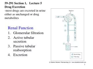
[AMJ 2019;12(2):63-70] Renal tubular acidosis in pediatrics: A review article Leena H Moshref, Rana H Moshref, and Osama Y Safdar King Abdulaziz University Hospital, Jeddah, Saudi Arabia consultants in the medical field. Key Words Renal tubular acidosis, constipation, hypokalaemia Review Please cite this paper as: Moshref LH, Moshref RH, Safdar OY. Renal tubular acidosis in pediatrics: A review article. AMJ 2019;12(2):63–70. https://doi.org/10.21767/AMJ.2018.3557 What this review adds: 1. What is known about this subject? Renal tubular acidosis is usually found in paediatrics and has various types. Treatment and early recognition is important to prevent growth stunting. 2. What new information is offered in this review? This review links many syndromes associated with renal tubular acidosis. It explores different aspects in management of different subtypes. 3. What are the implications for research, policy, or practice? Improvement in the management of renal tubular acidosis lies in early recognition and treatment, possibly gene therapy in the future. Corresponding Author: Leena Moshref King Abdulaziz University Hospital, Jeddah, Saudi Arabia Email: leenahatem0987@gmail.com ABSTRACT Background Renal tubular acidosis was first defined by the incapacity to lower urinary pH below 5.5, normal GFR associated with hyperchloremia and a normal plasma anion gap, and later the definition added tubular impairment in the bicarbonate reabsorption. Aims To provide an overview of types, diagnosis and management of renal tubular acidosis. The study focuses on syndromes found in Saudi Arabia. Methods This study is a review article about renal tubular acidosis. It is composed of an extensive research in PubMed, uptodate, BMJ, Cochrane. Results There are different hypothesis and genetic play a role in renal tubular acidosis. Treatment varies from each type, but with proper treatment promotes normal growth development. Conclusion This study is an overview of renal tubular acidosis. It provides a comprehensive summary to interns, residents and Introduction Renal tubular acidosis was first defined by the incapacity to lower urinary pH below 5.5, normal GFR associated with hyperchloremia and a normal plasma anion gap, and later the definition added tubular impairment in the bicarbonate reabsorption.1,2 Renal tubular acidosis (RTA) is related to a bunch of transport defects in bicarbonate reabsorption (HCO3 both.1 RTA can be divided to three types: distal RTA (type 1), proximal RTA (type 2), mixed (type 3), and hyperkalaemia RTA (type 4).3,4 RTA presents clinically with polyuria, constipation, growth failure, and muscular weakness, hypokalemic and hyperchloremic metabolic acidosis.2 Acid- base balance Extracellular fluid has free hydrogen ion concentration with concentration of 35 to 45nm/L, corresponding to pH of 7.35 to 7.45. In normal conditions, 0.8 to 1mmol/kg of non-volatile acids is generated daily mostly due to −), hydrogen ion secretion (H+), or 63
[AMJ 2019;12(2):63-70] 1- Idiopathic is associated with nephrolithiasis and/or nephrocalcinosis. Failure to thrive was the common presentation in infants, and later progresses to hypokalaemia further in adulthood. It is associated with gene mutations in kidney anion exchanger 1 (kAE1)5,6 caused by decrease in anion exchange thus hinder trafficking and regulation of V-type ATPase, venturing acidification of lysosomes.7 In a case report, down syndrome was associated hypothyroidism and renal acidosis, with antithyroid peroxidase antibodies positive and with treatment of hypothyroidism, symptoms of acidosis resolved.8 breakdown of dietary protein for energy. This is attenuated by the recycling of filtered bicarbonate and the excretion of protons with phosphate buffers (titratable acid) and ammonium. The filtered bicarbonate is reabsorbed by 70–80 per cent in the proximal tubule. The secretion of hydrogen ion in the proximal tubule is dependent on a Na-K exchanger, and is derived from basolateral sodium-potassium ATPase pumps, which preserves a low concentration of intracellular sodium and creates a chemical gradient for passive sodium entry from the tubular lumen. For 1 sodium absorbed, 1 hydrogen ion is secreted to the tubule, maintaining electro-neutrality. The bicarbonate combines with the hydrogen ion in the lumen to form carbonic acid, which later dissociates to CO2 and water. The carbon dioxide diffuses across the membrane and hydrates in the presence of carbonic anhydrase to form carbonic acid. This acid dissociates in the cell into hydrogen ion which diffuses to the basolateral membrane and bicarbonate which is exchanged with sodium across the brush border membrane. Ammonium ions generated by degradation of glutamite are transported from the proximal cell into the lumen in exchange with sodium ions. The kidney preserves the acid-base homeostasis by reabsorption of bicarbonate and excretion of acid. The acid excretion consists of ammonium and titratable acid ([TA+NH4 acidosis, the impairments involved are in acid excretion, bicarbonate reabsorption, generation of NH4+or buffer deficiency.2 The renal acid-base homeostasis mechanisms: (1) reabsorption of filtered HCO3−, which occurs mostly in the proximal convoluted tubule; and (2) excretion of fixed acids through the titration of buffers and excretion of ammonium, which occurs predominately in distal nephron.1 Classification of RTA Distal (Type 1): It is defined by impaired ability to acidify urine associated with hypokalaemia, low urinary NH4 hypocitraturia. Secondary to this defect, NH4 impaired. Hypothesis: a)Hydrogen ion secretory defect in collecting tubule. b)Gradient defect has increased back diffusion of H+ or H2CO3. c)Voltage dependent RTA which is inability to generate sufficient negative transepithelial difference in the distal nephron. d)Deficiency of carbonic anhydrase which is inability to produce intracellular bicarbonate results in failure to secrete hydrogen ion. e)Rate dependent RTA (2) which is divided to three types. A case series of periodic paralysis, otosclerosis and fluorosis, result from a mutation in anion exchanger 1 gene (AE1) codes band 3.9 A novel gene is discovered which is Atp6ap2 subunit of vH+ATPase known as prorenin receptor. Its suppression results in dilution of urine which is caused by lysosomal defect in vH+ATPase channel.10,11 2- Acquired cases as they are secondary to immune- mediated diseases. Most common association is with Sjogren’s syndrome and with kidney stones.12 3- Hereditary played a role in most RTA as they arose from hereditary hypercalciuria Hypocitraturia in most cases was due to potassium depletion and renal failure, but few were reported to occur as a primary defect in hereditary RTA. In a case series of Faqeih the renal tubular acidosis was associated with neurobehavioral nephrocalcinosis, facial features, small kidneys and short stature, but no genes are detected.12 Nephrocalcinosis is calcium deposition in renal medulla. Types include organic, iatrogenic and hereditary, and usually asymptomatic, if symptomatic, patients will present with inability to gain weight, blood in the urine and abdominal pain. It is associated with tubular acidosis, bratter and lowe syndromes, medullary sponge kidney, policalicosis, hypervitaminosis hypercalcuria, hypercalcemia hypoparathyroidism.12,13 Hemoglobinopathies due SLC4A1 encoding AE1, band 3, are causes of hereditary RTA. Patients with homozygous G701D/G701D or heterozygous SAO/G701D had reticulocytosis, which means compensated haemolysis. Both homozygous and heterozygous SLC4A1 mutations and hemoglobinopathy +] - HCO3). In renal tubular complication. depends on two impairment, + and + secretion is D, hyperoxaluria, and to mutations in gene 64
[AMJ 2019;12(2):63-70] affects the morphology of red cells and the degree of haemolytic anaemia, which is worsen by acidosis.14 Cases of transfusion-dependent severe haemolytic anaemia and complete distal renal tubular acidosis, dyserythropoiesis is caused by novel nonsense mutation c.1430C>A (p.Ser477X) in exon 12.15 Severe forms of inherited distal RTA present with vomiting and abstruse dehydration, growth restriction and vitamin D deficiency.13,16 Proximal (Type 2) It is caused by a defect of HCO3 associated with lower renal HCO3 normally (22–26mmol/L) in infants and adults.1 Because of the distal tubules ability to acidify is intact, the urinary pH can be less than 5.5.2 It characterized by bicarbonate leaving kidney tubule lumen as CO2 following sodium dependent H+ secretion via Na+/H+ exchanger isoforms or minimally vacuolar H+- ATPase. The transport requires both membrane bound carbonic anhydrase (CA) type 4 and intracellular CA-2.4 Hypothesis: a)Deficiency in pump secretion or function. b)Carbonic anhydrase deficiency. c)Structural damage to the luminal membrane2 Types: 1-Hereditary types include the ones mentioned below: A-Fanconi’s syndrome reabsorption in the proximal renal tubule affiliated with renal tubular acidosis and low phosphate in serum17 in SLC34A1 gene, presents with short stature, cataracts, central nervous system calcification, renal stones and hypercalcemia.18 Fanconi-Bickel syndrome missense mutation in SLC2A2,19 is a rare autosomal recessive disease caused by a deficiency of glucose transporter 2 (GLUT2), a member of the facilitative glucose transporter family.20 The most common type is cystinosis caused by CTNS mutation and is a lysosomal storage disorder.21 It usually responds to cysteamine.22 In 2 Palestinian patients from 2 families, homozygosity of genes were noted in p.R301X (C>T) in exon 7 of GLUT2 gene.20 It presents with fasting hypoglycaemia, post-prandial hyperglycaemia, delay in motor function, distended abdomen, unable to gain weight and renal tubular acidosis.23 B-Inborn error of metabolism including glucose-galactose malabsorption and congenital NBCe1A deficiency, cystinosis and tyrosinemia as example. Glucose-galactose malabsorption is caused by mutations in the gene that resides on the distal q arm of chromosome 22 coding for the intestinal brush border sodium-glucose co-transporter (SGLT1). It is manifested by early onset of profuse osmotic watery diuresis and can lead to hypernatremia, metabolic acidosis and mortality if left untreated.24 Congenital NBCe1A deficiency with SLC4A4 mutation manifests with severe proximal renal tubular acidosis and extrarenal symptoms, such as developmental delay, glaucoma, cataract, intellectual disability and band keratopathy.25 It usually has no treatment.26 Tyrosinemia type I results fumarylacetoacetase (FAH), dysfunction and renal tubular dysfunction associated with growth failure and rickets. Screening is done by urine and blood succinylacetone.27 2-Acquired isolated proximal tubule is caused by a defect of bicarbonate reabsorption, in which mutations are found in sodium bicarbonate co- transporter (NBCe1/SLC4A4).28 A-Carbonic anhydrase deficiency secondary to drugs, Diamox, Sulfanilamide, genetically transmitted (Russell), Idiopathic (Donkerwolke), caused by homozygous mutations chromosome 8q22. The commonest gene mutation is loss of splice donor site in intron 2 of CA2 in Saudi Arabia. It presents as paralysis associated with fractures, renal stones, growth retardation, osteopetrosis and mental retardation.29 B-Idiopathic cases, such as, York-Yendt, transient in infants usually present with short stature, vomiting, diarrhoea and feeding difficulties in severe cases.30 Case report of distal renal tubular acidosis with hearing loss confirmed by audiometry at 2 years. ATP6V0A4 was excluded from genetic causation by intragenic SNP linkage analysis, but ATP6V1B1 completely failed to PCR-amplify in the patient, suggesting a genomic deletion.31 Combined (Type 3) It is caused by recessive mutation in the CA-2 gene on 8q22, which encodes carbonic anhydrase type 2. It exhibits both impaired proximal HCO3 impaired distal acidification.4 − reabsorption and is − threshold, which is from presents deficiency with of liver in CA2 located on is defined as impaired - reabsorption and 65
[AMJ 2019;12(2):63-70] Bicarbonate wasting goes away after adolescence or after rapid growth spurts. It is considered a paediatric variant of type 1 since it is in infants and children.30 Hyperkalaemia (Type 4) The commonest type of RTA is type 4. It results from impaired ammoniagenesis and is characterized by the ability to normally acidify the urine after an acid load results in low urine NH4 by aldosterone deficiency or defective signalling; like, adrenal failure or pseudohypoaldosteronism (PHA) due to defects in the mineralocorticoid receptor or the epithelial Na+ channel, drugs (e.g., potassium sparing diuretics, trimethoprim, cyclo- oxygenase inhibitors, angiotensin inhibitors).1,4 Hypothesis: a)Selective destruction of the juxtaglomerular cells of kidney. b)Decreased sympathetic innervation of the juxtaglomerular apparatus. c)Decreased production of prostacyclin proceeds from decrease in the renin-angiotensin production. d)Hypoaldosteronism. Five types: Type 1 & 2 is a consequence of primary hormonal deficiency of renin/ aldosterone thus affecting distal convoluted tubule. Type 1 is defined as decreased urinary aldosterone, increased plasma renin and salt wasting, resulting in acidosis, hyperkalaemia, and is usually linked to Addison disease, isolated primary hyperaldosteronism, or congenital adrenal hyperplasia. Type 2 is known as hyporeninemic hypoaldosteronism, and is described as renal renin impairment resulting from chronic interstitial damage. To note, clinical salt wasting doesn’t prevail, probably due to constant azotaemia. Type 3, 4 & 5 are disorders of renal tubules. Type 3 (adolescent hyperkalemic syndrome) or (renal chloride shunt) is defined by hypervolemia, hypoaldosteronism hypertension. Surprisingly, hypervolemia and secondary suppression of aldosterone result from excessive chloride reabsorption in distal convoluted tubule (Table 1). Type 4 (pseudohypoaldosteronism) in infants is defined by acidosis, hyperkalaemia, sodium chloride wasting and elevated 24- urinary aldosterone excretion. It is caused by unresponsive distal tubules to aldosterone; such as, organ failure. However, type 4 rarely occurs due to adequate treatment with sodium bicarbonate and sodium chloride. Type 5 (early childhood type 4 RTA) is the commonest type of type 4 RTA, and is defined by hyperchloremic acidosis, aciduria and hyperkalaemia, with no clinical salt wasting. It is proposed that type 5 is a cause of end organ resistance to 2 out of 3 aldosterone dependent functions (potassium and hydrogen reduced, sodium is normal). Children with type 5 usually end up short, stunted and fail to thrive. There are two factors that predispose to renal stone formation in subtype 5 of type 4 RTA, which are urinary calcium and citrate excretion. During chronic acidosis, the mobilization of bone buffers is increased, and in children with type 1 and type 2 RTA, urinary calcium excretion rises. However, in children with subtype 5 of type 4 RTA have no rise in urinary calcium excretion during chronic acidosis. Consequently, untreated, chronically acidotic patients with subtype 5 of type 4 RTA, nephrocalcinosis does not occur because of calcium hyperabsorption and citrate excretion in urine.30 Hyperkalemic renal tubular acidosis caused by calcineurin inhibitors (Figures 1 and 2).32 Diagnosis Figure 1: RTA differentiated by hyperchloremic acidosis33 +, which is probably caused by the hyperkalaemia, or converting enzyme and 66
[AMJ 2019;12(2):63-70] Figure 2: RTA differentiated by hypokalemic acidosis16 can be avoided by giving diuretics and diet high in salt. Other modalities of treatment include potassium exchange resin and alkali, may also be necessary.35 Metabolic acidosis if corrected, will lead to normal growth rates in children, decreasing the rates of nephrocalcinosis and osteopenia.34 If hypokalaemia is present, potassium citrate (60–80mEq. daily) for three months nephrolithiasis.34 A case of Sjogren’s syndrome refractory type 1 renal tubular acidosis was treated by sodium bicarbonate 15.5mEq four times a day, potassium chloride and amiloride 10mg/day.11 nephrocalcinosis underlying causes, symptoms will resolve.12 Latest research precludes that disruption of the COPI- kAE1 G701D interaction could be a therapeutic strategy to treat dRTA caused by this mutant.6 Type 2 RTA (Proximal) Proximal RTA has a difficult approach in comparison to distal RTA, for serum bicarbonate increases the filtered bicarbonate causing diuresis; therefore, an increase bicarbonate is needed in 10–15meq of alkali/kg/day with the addition of potassium salt (usually potassium citrate)34 in cases Proximal RTA with Fanconi syndrome usually responds to Vitamin D replacement.36 Dietary salt restriction and chlorothiazide therapy can be useful if alkali is ineffective or not well tolerated in massive amounts.1,30 Congenital NBCe1A deficiency has no treatments available at the moment, but in-vitro studies showed that aminoglycoside antibiotic is associated with suppression of the genes.26 Cystinosis mainstay treatment is oral cysteamine (Cystagon). Aminothiol can lower intracellular cystine content by 95 per cent, and has proven efficacy in delaying renal glomerular growth and preventing hypothyroidism.22 Supplementary treatment involves supplementations phosphate replacement and vitamin D supplements.37 Tyrosinemia responds to nitisinone and a low-tyrosine diet as it prolongs survival, enhance normal growth, can help to reduce With treatment of Confirmatory tests Table 1: Confirmatory Tests16 Proximal RTA Distal RTA Hyperkalemic RTA Urinary measurement of acid and bicarbonate Decline of serum HCO3 and pH Inability to excrete H - - High or low, depends on reason for hypoaldosteronism Serrum aldosterone - - rickets.30 of + loading NH4 test: urinary pH will not decrease as expected. Urine anion gap is used as a replacement marker of NH4 secretion. - HCO3 loading test: urinary HCO3 will rise following HCO3 infusion because of impaired reabsorption. - Loading test (Side bar) - - + Management Type 1 RTA (Distal): Alkali has to be compensated by the amount of retained hydrogen and loss urinary bicarbonate calculated by Henderson-Hasselbalch equation, which will result in achieving normal serum bicarbonate concentration (22–24meq/L). Children may require 4-8meq/kg per day in divided doses because they often have a higher fixed urine pH.34 Mineralocorticoid therapy is beneficial in patients with aldosterone deficiency. Giving steroid without increasing distal sodium delivery can cause heart failure. Heart failure deterioration, enhancing of citrate, 67
[AMJ 2019;12(2):63-70] improve liver function, correct acidosis, and improve secondary rickets.37 Liver transplant used in refractory cases of proximal RTA including Fanconi and tyrosenemia.23,27 Kidney transplant as a definitive treatment for cystenosis.37 Type 3 RTA Alkali therapy can treat osteopenia and induce normal somatic growth, even after severe stunting, but surprisingly only when given in amounts that sustain full correction of acidosis.38 Type 4 RTA Treatment depends on the underlying cause. Potassium- retaining drugs should Hypoaldosteronism caused by adrenal failure may respond to fludrocortisone doses. hypoaldosteronism, fludrocortisone with the addition of loop diuretic help to minimize the risk of extracellular fluid volume expansion. Alkali supplements (1.5–2mmol/kg per 24 h) are required.39,40 Conclusion This study is an overview of renal tubular acidosis. It provides a comprehensive summary to interns, residents and consultants in the medical field. This review links many syndromes associated with renal tubular acidosis. It explores different aspects in management of different subtypes. Improvement in the management of renal tubular acidosis lies in early recognition and treatment, possibly gene therapy in the future. Membrane 10.1080/09687688.2018.1451662. 6.Duangtum N, Junking M, Phadngam S, et al. γ-COPI mediates the retention of kAE1 G701D protein in Golgi apparatus - a mechanistic explanation of distal renal tubular acidosis associated with the G701D mutation. Biochemical J. 2017;474(15):2573–2584. 10.1042/BCJ20170088. 7.Mumtaz R, Trepiccione F, Hennings JC, et al. Intercalated Cell Depletion and Vacuolar H+-ATPase Mistargeting in an Ae1 R607H Knockin Model. J Am Society of Nephrology. 2017;28(5):1507–1520. doi: 10.1681/ASN.2016020169. 8.Spahiu L, Jashari H, Mulliqi-Kotori V, et al. Hashimoto Thyroiditis and Nephrocalcinosis in a Child with Down Syndrome. Acta 2016;24(2):143–5. doi: 10.5455/aim.2016.24.143-145. 9.Vithanage JP, Ekanayake M. A case of distal renal tubular acidosis, Southeast Asian ovalocytosis and possible fluorosis. Journal. 2009;54(1):19–20. 10.4038/cmj.v54i1.469. 10.Trepiccione F, Prosperi F, de la Motte L, et al. New Findings on the Pathogenesis of Distal Renal Tubular Acidosis. Kidney Diseases. 2017;3(3):98–105. doi: 10.1159/000478781. 11.Oguejiofor P, Chow R, Yim K, et al. Successful Management of Refractory Type 1 Renal Tubular Acidosis with Amiloride. Nephrology. 2017;2017:1–3. 10.1155/2017/8596169. 12.Faqeih E, Al-Akash S, Sakati N, et al. Four siblings with distal renal tubular acidosis and nephrocalcinosis, neurobehavioral impairment, short stature, and distinctive facial appearance: A possible new autosomal recessive syndrome. American Journal of Medical Genetics Part A. 2007;143A(17):1951–1957. doi: 10.1002/ajmg.a.31898. 13.Pitukweerakul S, Prachuapthunyachart S. Bilateral Nephrocalcinosis in Primary Distal Renal Tubular Acidosis. Journal of General Internal Medicine. 2016;31(10):1261–1261. doi: 10.1007/s11606-016- 3697-z. 14.Khositseth S, Sirikanaerat A, Khoprasert S, et al. Hematological abnormalities in patients with distal renal tubular acidosis and hemoglobinopathies. American Journal of Hematology. 2008;83(6):465–471. doi: 10.1002/ajh.21151. 15.Kager L, Bruce L, Zeitlhofer P, et al. Band 3 nullVIENNA, a novel homozygousSLC4A1p.Ser477X variant causing severe hemolytic anemia, dyserythropoiesis and Biology. 2017;34(1-2):50–64. doi: doi: always be withdrawn. Informatica Medica. As for hyporeninemic Ceylon Medical doi: Case Reports in doi: References 1.Rodriguez Soriano J. Renal Tubular Acidosis: The Clinical Entity. Journal of the American Society of Nephrology. 2002;13(8):2160–2170. doi: 10.1097/01.asn.0000023430.92674.e5. 2.Saborio P, Krieg RJ, Ahmad TM, et al. Renal tubular acidosis in children. Saudi Journal of Kidney Diseases and Transplantation. 1997;8(3):235–246. PMID:18417801. 3.Rodriguez-Soriano J, Vallo A. Renal tubular acidosis. Pediatric Nephrology. 10.1007/bf00857675. 4.Ring T, Frische Clinical review: Renal tubular acidosis- a physicochemicalapproach Critical Care. 2005;9(6):573. doi: 10.1186/cc3802. 5.Almomani E, Touret N, Cordat E. Adaptor protein 1 B mu subunit does not contribute to the recycling of kAE1 protein in polarized renal epithelial cells. Molecular 1990;4(3):268–275. doi: S, Nielsen S. 68
[AMJ 2019;12(2):63-70] complete distal renal tubular acidosis. Pediatric Blood & Cancer. 2016;64(3):e26227. doi: 10.1002/pbc.26227. 16.Yaxley J, Pirrone C. Review of the Diagnostic Evaluation of Renal Tubular Acidosis. Ochsner Journal. 2016;16(4):525– 530. PMID:27999512 PMCID:PMC5158160. 17.Gutierrez J, Zurita M, Zurita L. Sjögren’s Syndrome Associated with Fanconi’s Syndrome and Osteomalacia. American Journal of Case Reports. 2018;19:392–396. doi: 10.12659/ajcr.907503. 18.Fearn A, Allison B, Rice S, et al. Clinical, biochemical, and pathophysiological analysis of SLC34A1 Physiological Reports. 10.14814/phy2.13715. 19.Khandelwal P, Sinha A, Jain V, et al. Fanconi syndrome and neonatal diabetes: phenotypic heterogeneity in patients with GLUT2 defects. CEN Case Reports. 2017;7(1):1–4. doi: 10.1007/s13730-017-0278-x. 20.Dweikat I, Alawneh I, Bahar S, et al. Fanconi-Bickel syndrome in two Palestinian children: marked phenotypic variability with identical mutation. BMC Research Notes. 2016;9(1). doi: 10.1186/s13104-016-2184-2. 21.Elmonem M, Veys K, Soliman N, et al. Cystinosis: a review. Orphanet Journal of Rare Diseases. 2016;11(1). doi: 10.1186/s13023-016-0426-y. 22.Gahl W. Early oral cysteamine therapy for nephropathic cystinosis. Eur J Pediatrics. 2003;162(0):S38–S41. doi: 10.1007/s00431-003-1349-x. 23.Kehar M, Bijarnia S, Ellard S, et al. Fanconi-Bickel Syndrome - Mutation in SLC2A2 Gene. The Indian Journal of Pediatrics. 2014;81(11):1237–1239. 10.1007/s12098-014-1487-3. 24.El-Naggar W, Balfe J, Barbar M, et al. Nephrocalcinosis in glucose-galactose malabsorption, association with renal tubular acidosis. Pediatric Nephrology. 2005;20(9):1336– 1339. doi: 10.1007/s00467-005-1885-x. 25.Horita S, Simsek E, Simsek T, et al. SLC4A4 compound heterozygous mutations in exon–intron boundary regions presenting with severe proximal renal tubular acidosis and extrarenal symptoms coexisting with Turner’s syndrome: a case report. BMC Medical Genetics. 2018;19(1). doi: 10.1186/s12881-018-0612-y. 1993-2018. Available https://www.ncbi.nlm.nih.gov/books/NBK1515/ 28.Kari J, El Desoky S, Singh A, et al. The Case | Renal tubular acidosis and eye findings. Kidney International. 2014;86(1):217–218. doi: 10.1038/ki.2013.320. 29.Alsharidi A, Al-Hamed M, Alsuwaida A. Carbonic anhydrase II deficiency: report of a novel mutation. CEN Case Reports. 10.1007/s13730-015-0205-y. 30.McSherry P. Renal tubular acidosis in childhood. Kidney International. 10.1038/ki.1981.213. 31.Norgett E, Yii A, Blake-Palmer K, Sharifian M, et al. A role for VAX2 in correct retinal function revealed by a novel genomic deletion at 2p13.3 causing distal Renal Tubular Acidosis: case report. BMC Medical Genetics. 2015;16(1). doi: 10.1186/s12881-015-0182-1. 32.Lin W, Mou L, Tu H, et al. Clinical analysis of hyperkalemic renal tubular acidosis caused by calcineurin inhibitors in solid organ transplant recipients. Journal of Therapeutics. 2016;42(1):122–124. 10.1111/jcpt.12485. 33.Muzalef A, Alshehri M, Al-Abidi A, et al. Marble brain disease in two Saudi Arabian siblings. Annals of Tropical Paediatrics. 10.1179/146532805X58175. 34.Aronson P, Giebisch G. Effects of pH on Potassium: New Explanations for Old Observations. Journal of the American Society of Nephrology. 2011;22(11):1981– 1989. doi: 10.1681/ASN.2011040414. 35.Kurtzman N. Disorders of distal acidification. Kidney International. 1990;38(4):720–727. 10.1038/ki.1990.264. 36.Ali S, Tariq M. Successful treatment of proximal renal tubular acidosis and Fanconi syndrome with vitamin D replacement. Saudi J Transplantation. 2016;27(4):812. doi: 10.4103/1319- 2442.185270. 37.Nesterova G, Gahl WA. Cystinosis. 2001 Mar 22 [Updated 2017 Dec 7]. In: Adam MP, Ardinger HH, Pagon RA, et al., editors. GeneReviews® [Internet]. Seattle (WA): University of Washington, Seattle; 1993- 2018. Available https://www.ncbi.nlm.nih.gov/books/NBK1400/ 38.Morris R. Alkali Therapy In Renal Tubular Acidosis: Who Needs It?. Journal of the American Society of Nephrology. 2002;13(8):2186–2188. 10.1097/01.asn.0000027973.07189.00. 39.Sebastian A, Schambelan M, Sutton J. Amelioration of Hyperchloremic Acidosis with Furosemide Therapy in from: 2015;5(1):108–112. doi: mutations. 1981;20(6):799–809. doi: 2018;6(12):e13715. doi: Clinical Pharmacy and doi: 2005;25(3):213–218. doi: doi: doi: Kidney Diseases and 26.Azimov R, Abuladze N, Sassani P, et al. G418-mediated ribosomal read-through of a nonsense mutation causing autosomal recessive proximal renal tubular acidosis. American Journal of Physiology-Renal 2008;295(3):F633–F641. 10.1152/ajprenal.00015.2008. 27.Sniderman King L, Trahms C, Scott CR. Tyrosinemia Type I. 2006 Jul 24 [Updated 2017 May 25]. In: Adam MP, Ardinger HH, Pagon RA, et al., editors. GeneReviews® [Internet]. Seattle (WA): University of Washington, Seattle; from: Physiology. doi: doi: 69
[AMJ 2019;12(2):63-70] Patients with Chronic Renal Insufficiency and Type 4 Renal Tubular Acidosis. American Journal of Nephrology. 1984;4(5):287–300. doi: 10.1159/000166827. 40.Sebastian A, Schambelan M, Lindenfeild S, et al. Amelioration of Metabolic Acidosis with Fludrocortisone Therapy in Hyporeninemic Hypoaldosteronism. New England Journal of Medicine. 1977;297(11):576–583. doi: 10.1056/NEJM197709152971104. PEER REVIEW Not commissioned. Externally peer reviewed. CONFLICTS OF INTEREST The authors declare that they have no competing interests. FUNDING None ETHICS COMMITTEE APPROVAL There is no ethical approval as it is a systemic review. 70