Download
1 / 88
880 likes | 882 Views
This chapter covers the cell theory, cellular organization, plasma membrane, cytoplasm, organelles, nucleus, protein synthesis, and cell division.

E N D
Chapter 3 Cells and Tissues Part 1: Cells
Outline • The Cell Theory • Cellular Organization (The Parts of a Cell) • Plasma Membrane • Transport across the plasma membrane • Cytoplasm • Cytosol • Organelles • Nucleus • Protein Synthesis • Cell Division • Mitosis
The Cell Theory • Let’s face it, life is totally cellular (this does not refer to the mobile phone) • The cell theory states: • Cells are the basic unit of life • All living things are made up of cells • New cells arise only from preexisting cells
Table 3.1 Parts of the Cell: Structure and Function (1 of 5).
Table 3.1 Parts of the Cell: Structure and Function (2 of 5).
Table 3.1 Parts of the Cell: Structure and Function (3 of 5).
Table 3.1 Parts of the Cell: Structure and Function (4 of 5).
Table 3.1 Parts of the Cell: Structure and Function (5 of 5).
Cellular Organization • Plasma membrane surrounds the cell and regulates entrance and exit of substances • Selective barrier • Key role in communication between cells • Key role in communication of cells with the external environment • Cytoplasm is the portion of the cell between the nucleus and plasma membrane • Cytosol: fluid portion of cytoplasm • Consistency: semifluid gel, like wet Jello • Organelles: small membranous structures each with a specific function • The Nucleus- storage of genetic information
Cellular Organization (Cont.) • Nucleus is the centrally located organelle containing chromosomes and is the control center of the cell
Chromatin Nuclear envelope Smoothendoplasmicreticulum Nucleolus Nucleus Plasmamembrane Cytosol Lysosome Figure 3.4 Structure of the generalized cell. Mitochondrion Roughendoplasmicreticulum Centrioles Ribosomes Golgiapparatus Secretion beingreleased from cellby exocytosis Microtubule Peroxisome Intermediatefilaments
Plasma Membrane • Plasma membrane is a phospholipid bilayer with attached or embedded proteins • Fluid mosaic model • Sea of fluid lipids with mosaic of different proteins • Phospholipids: polar head and non-polar tails • Form spherical bilayer when placed in water • Cholesterol • Glycolipids • Membrane Fluidity: critical for interactions of membrane proteins and allows cellular process e.g. movement, growth, division, secretion • Dependent on • Number of double bonds in the fatty acid tails of the lipids • Amount of cholesterol
Hydrophilic: Love water Extracellular fluid(watery environment) Glycoprotein Glycolipid Cholesterol Figure 3.2 Structure of the plasma membrane. Sugargroup Polar headsof phospholipidmolecules Bimolecularlipid layercontainingproteins Channel Nonpolar tailsof phospholipidmolecules Proteins Filaments ofcytoskeleton Cytoplasm(watery environment) Hydrophobic: Hate water
Plasma Membrane (Cont.) • Plasma membrane is selectively permeable, and regulates movement of molecules and ions across the cell membrane due to nature of the phospholipids • Plasma membrane proteins form receptors, conductors, or enzymes in metabolic reactions
Extracellular fluid(watery environment) Glycoprotein Glycolipid Cholesterol Figure 3.2 Structure of the plasma membrane. Sugargroup Polar headsof phospholipidmolecules Bimolecularlipid layercontainingproteins Channel Nonpolar tailsof phospholipidmolecules Proteins Filaments ofcytoskeleton Cytoplasm(watery environment)
The Plasma Membrane • Examples of different membrane proteins include • Ion channels • Carriers • Receptors
The Plasma Membrane • Examples of different membrane proteins include • Enzymes • Linkers • Cell identity markers
Plasma Membrane (Cont.) • Transport across the plasma membrane- Two Types • Passive Processes- don’t require ATP • Active Processes- require ATP • Passive processes • Simple diffusion: movement of molecule down concentration gradient • Facilitated diffusion • Channel-mediated • Carrier-mediated • Osmosis: diffusion of water down it’s concentration gradient • Water moves through bilayer • Water moves through aquaporins • Concept of Tonicity • Must have balance of solutes and water due to selective permeability of membrane to solutes
Plasma Membrane (Cont.) • Transport across the plasma membrane (Cont.) • Passive Processes (Cont.) • Simple Diffusion is the random movement of molecules from an area of higher concentration to an area of lower concentration until they are equally distributed
Plasma Membrane (Cont.) • Transport across the plasma membrane (Cont.) • Passive Processes (Cont.) • Facilitated diffusion • Channel-mediated • Carrier-mediated • Osmosis: diffusion of water down it’s concentration gradient • Water moves through bilayer • Water moves through aquaporins
Small lipid-insolublesolutes Extracellularfluid Lipid-insolublesolutes Watermolecules Lipid-solublesolutes Figure 3.10 Diffusion through the plasma membrane. Lipidbilayer Cytoplasm (b) Carrier-mediatedfacilitated diffusion viaprotein carrier specific forone chemical; binding ofsubstrate causes shapechange in transport protein (c) Channel-mediatedfacilitateddiffusionthrough achannel protein;mostly ions,selected onbasis ofsize and charge (a) Simplediffusionof fat-solublemoleculesdirectlythrough thephospholipidbilayer (d) Osmosis,diffusionof water through aspecific channelprotein (aquaporin)or through the lipid bilayer
Plasma Membrane (Cont.) • Transport across the plasma membrane (Cont.) • Passive Processes (Cont.) • Osmosis: diffusion of water down it’s concentration gradient • Water moves through bilayer • Water moves through aquaporins • Osmosis is the random movement of water from an area of higher concentration of water to an area of lower concentration of water • Important due to the selective permeability of membrane to solutes • Leads to the concept of tonicity • Tonicity: the ability of a solution to change the water content of a cell due to selective permeability of the membrane and solutes in the solution
Principle of Osmosis Applied pressure = osmotic pressure Left arm Right arm Volumes equal Water molecule Osmosis Osmosis Selectively permeable membrane Solute molecule Movement due to hydrostatic pressure (c) Restoring starting conditions (b) Equilibrium (a) Starting conditions
Isotonic solution Hypotonic solution Hypertonic solution (a) Illustrations showing direction of water movement SEM Normal RBC shape RBC undergoes hemolysis RBC undergoes crenation (b) Scanning electron micrographs (all 15,000x)
Plasma Membrane (Cont.) • Transport across the plasma membrane (Cont.) • Active processes:need to utilize ATP to drive molecular movement against concentration gradient • Active Transport: Carrier-mediated transport of molecules by proteins in plasma membrane • Vesicular Transport: transport in vesicles, moving substances into or out of the cell • Exocytosis: vesicular transport out of the cell • Endocytosis: vesicular transport into the cell • Phagocytosis- uptake of bacteria or dead cells • Receptor-mediated- specific binding to cell surface protein before uptake • Pinocytosis- uptake of fluid (cell drinking)
Extracellular fluid Na+ Na+ K+ 2 3 1 3 2 1 Na+-K+ pump Na+ Figure 3.11 Operation of the sodium-potassium pump, a solute pump. Na+ Na+ K+ P K+ P Na+ ATP K+ ADP Loss of phosphaterestores the originalconformation of the pumpprotein. K+ is released to thecytoplasm, and Na+ sites areready to bind Na+ again; the cycle repeats. Binding of cytoplasmic Na+to the pump protein stimulatesphosphorylation by ATP, which causes the pump protein tochange its shape. The shape change expelsNa+ to the outside. ExtracellularK+ binds, causing release of thephosphate group. Sodium-Potassium Pump Cytoplasm
Plasma Membrane (Cont.) • Transport across the plasma membrane (Cont.) • Active processes (Cont.) • Vesicular Transport: transport in vesicles, moving substances into or out of the cell • Exocytosis: vesicular transport out of the cell
PlasmamembraneSNARE(t-SNARE) Extracellular fluid 2 The membrane-bound vesiclemigrates to the plasma membrane. 1 VesicleSNARE(v-SNARE) 3 Moleculeto besecreted Figure 3.12 Exocytosis. Secretoryvesicle Cytoplasm Fusion pore formed There, v-SNAREsbind with t-SNAREs,the vesicle andplasma membranefuse, and a poreopens up. Fused SNAREs Vesicle contentsare released to thecell exterior. (b) Electron micrograph of asecretory vesicle inexocytosis (190,000×) (a) The process of exocytosis
Plasma Membrane (Cont.) • Transport across the plasma membrane (Cont.) • Active processes (Cont.) • Vesicular Transport: transport in vesicles, moving substances into or out of the cell • Endocytosis: vesicular transport into the cell • Phagocytosis- uptake of bacteria or dead cells • Receptor-mediated- specific binding to cell surface protein before uptake • Pinocytosis- uptake of fluid (cell drinking)
Extracellularfluid Cytoplasm Extracellularfluid Plasmamembrane Cytosol Bacteriumor otherparticle 2 3 1 Vesicle Lysosome • Vesicle fusingwith lysosomefor digestion Release ofcontents tocytosol Figure 3.13 Events and types of endocytosis. • Transport to plasmamembrane and exocytosisof vesicle contents Pseudopod Phagocytosis (b) Detached vesicle Ingestedsubstance Membranereceptor • Membranes and receptors(if present) recycled to plasmamembrane Pit Pinocytosis Receptor-mediated (a) (c)
Cellular Organization • Plasma membrane surrounds the cell and regulates entrance and exit of substances • Selective barrier • Key role in communication between cells • Key role in communication of cells with the external environment • Cytoplasm is the portion of the cell between the nucleus and plasma membrane • Cytosol: fluid portion of cytoplasm • Consistency: semifluid gel, like wet Jello • Organelles: small membranous structures each with a specific function • The Nucleus- storage of genetic information
Cytoplasm • Cytoplasm is the portion of the cell between the nucleus and plasma membrane • Cytosol: fluid portion of cytoplasm • Consistency: semifluid gel, like wet Jello • The Cytoskeleton • Organelles: small membranous structures each with a specific function • Centrosome- contains two centrioles • Cilia & Flagella • Ribosomes • Endoplasmic Reticulum • Golgi Complex • Lysosomes • Mitochondria
The Cytosol • Cytoskeleton is a network of interconnected filaments and microtubules in the cytoplasm that maintain cell shape • The cytoskeleton is formed of several types of filamentous structures that give the cell its shape and organelles the ability to move about the cell • Microfilaments: actin & myosin; provide movement, mechanical support • Intermediate filaments: keratin; stabilize organelles, attach cells to one another • Microtubules: tubulin; movement of organelles, cilia & flagella
(b) Intermediate filaments (c) Microtubules (a) Microfilaments Tubulin subunits Fibrous subunits Actin subunit Figure 3.7 Cytoskeletal elements support the cell and help to generate movement. 25 nm 10 nm 7 nm Intermediate filaments formthe purple networksurrounding the pink nucleus. Microfilaments form the bluebatlike network. Microtubules appear as goldnetworks surrounding thecells’ pink nuclei.
Organelles • Centrosomes: contains two centrioles made of tubulin; organizing center for mitotic spindle during cell division & for microtubule formation in non-dividing cells • Cilia & Flagella • Cilia: microtubules; move fluids along a cell’s surface • Flagella: microtubules; moves an entire cell, e.g. sperm
Chromatin Nuclear envelope Smoothendoplasmicreticulum Nucleolus Nucleus Plasmamembrane Cytosol Lysosome Figure 3.4 Structure of the generalized cell. Mitochondrion Roughendoplasmicreticulum Centrioles Ribosomes Golgiapparatus Secretion beingreleased from cellby exocytosis Microtubule Peroxisome Intermediatefilaments
Organelles • Centrosome - located near the nucleus, consists of two centrioles and pericentriolar material
Organelles • Cilia - short, hair-like projections from the cell surface, move fluids along a cell surface • Flagella - longer than cilia, move an entire cell; only example is the sperm cell’s tail
Organelles (Cont.) • Ribosomes • Composed of multiple proteins and ribosomal RNA (rRNA) • Function in protein synthesis • Ribosomes on endoplasmic reticulum: synthesize proteins destined for insertion into plasma membrane or secretion from cell • Ribosomes free in cytosol: synthesize proteins used in the cytosol
Chromatin Nuclear envelope Smoothendoplasmicreticulum Nucleolus Nucleus Plasmamembrane Cytosol Lysosome Figure 3.4 Structure of the generalized cell. Mitochondrion Roughendoplasmicreticulum Centrioles Ribosomes Golgiapparatus Secretion beingreleased from cellby exocytosis Microtubule Peroxisome Intermediatefilaments
Ribosome 1 1 2 3 4 2 3 4 mRNA Rough ER • As the protein is synthesized on theribosome, it migrates into the rough ERcistern. Figure 3.5 Synthesis and export of a protein by the rough ER. • In the cistern, the protein folds into itsfunctional shape. Short sugar chains may beattached to the protein (forming aglycoprotein). Protein Transportvesicle buds off • The protein is packaged in a tinymembranous sac called a transport vesicle. • The transport vesicle buds from therough ER and travels to the Golgi apparatusfor further processing. Protein insidetransport vesicle
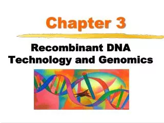
Organelles (Cont.) • Endoplasmic Reticulum(ER) • System of membranous channels and saccules • Rough ER: studded with ribosomes • Synthesizes glycoproteins, phospholipids, to be transferred into organelles, plasma membrane, or secreted • Smooth ER: no ribosomes • Synthesizes fatty acids, steroids, stores calcium for muscle contraction in muscle cells
Chromatin Nuclear envelope Smoothendoplasmicreticulum Nucleolus Nucleus Plasmamembrane Cytosol Lysosome Figure 3.4 Structure of the generalized cell. Mitochondrion Roughendoplasmicreticulum Centrioles Ribosomes Golgiapparatus Secretion beingreleased from cellby exocytosis Microtubule Peroxisome Intermediatefilaments
Ribosome 1 1 2 3 4 2 3 4 mRNA Rough ER • As the protein is synthesized on theribosome, it migrates into the rough ERcistern. Figure 3.5 Synthesis and export of a protein by the rough ER. • In the cistern, the protein folds into itsfunctional shape. Short sugar chains may beattached to the protein (forming aglycoprotein). Protein Transportvesicle buds off • The protein is packaged in a tinymembranous sac called a transport vesicle. • The transport vesicle buds from therough ER and travels to the Golgi apparatusfor further processing. Protein insidetransport vesicle
Organelles (Cont.) • The Golgi Complex • Consists of a stack of three to twenty curved cisternae, along with vesicles • Modifies, sorts, packages, transports proteins that bud from the rough ER • Forms secretory vesicles for ferrying molecules • Destined for exocytosis • Destined for plasma membrane • Destined for other organelles; e.g. lysosomes
Rough ER Cisterns Proteins in cisterns Lysosome fuseswith ingestedsubstances. Membrane Transportvesicle Figure 3.6 Role of the Golgi apparatus in packaging the products of the rough ER. Golgi vesicle containingdigestive enzymesbecomes a lysosome. Pathway 3 Pathway 2 Golgi vesicle containingmembrane componentsfuses with the plasmamembrane and isincorporated into it. Golgiapparatus Secretory vesicles Pathway 1 Proteins Golgi vesicle containingproteins to be secretedbecomes a secretoryvesicle. Plasma membrane Secretion byexocytosis Extracellular fluid
Synthesized protein Ribosome Entry face cisterna Transport vesicle 1 2 Medial cisterna 3 Exit face cisterna 9 4 Transport vesicle (to lysosome) 8 6 Rough ER Transfer vesicle Membrane vesicle 4 Proteins in vesicle membrane merge with plasma membrane 7 5 Transfer vesicle Secretory vesicle Proteins exported from cell by exocytosis Plasma membrane