Download
1 / 32
320 likes | 526 Views
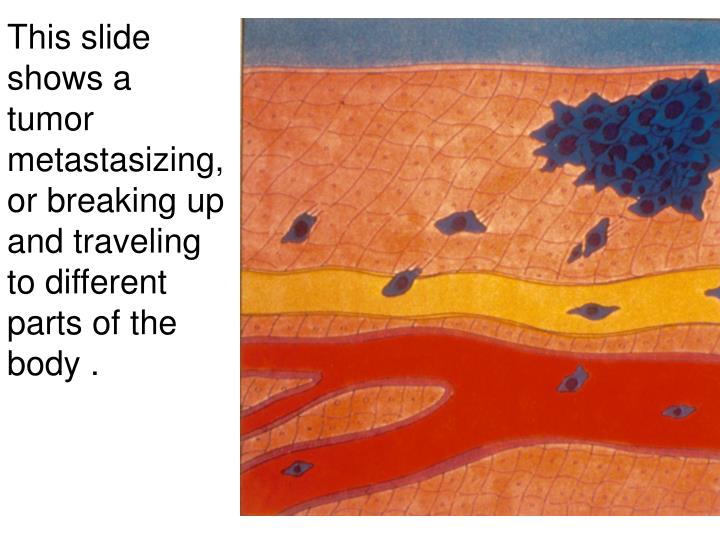
This slide shows a tumor metastasizing, or breaking up and traveling to different parts of the body. Basal Cell Carcinoma – the most common type - up to 90% of all skin cancers are this.

E N D
This slide shows a tumor metastasizing, or breaking up and traveling to different parts of the body .
Basal Cell Carcinoma – the most common type - up to 90% of all skin cancers are this.
This is another basal cell carcinoma. They typically start as a shiny bump, then slowly grow and form disfiguring masses. They seldom metastasize. Removal is done surgically, and only require radiation in tough to treat cases.
When left untreated, basal cell carcinomas can cause great disfigurement. Surgical removal will leave large depressions in the skin’s surface.
The dark area in the center of this circle is a squamous cell carcinoma. After surgical removal, this patient will receive radiation treatment. Between 1500 and 2000 people die from this type of cancer each year.
This girl has a squamous cell carcinoma. People with high melanin content are not immune to skin cancers!
This is an example of melanoma or malignant melanoma. This is the most dangerous form, and also the most rare.
The ABCD Rule of Melanoma • A = Asymetry – one half of the mole does not match the other • B = Border Irregularity – the edges of the mole are uneven, notched or blurred • C = Color – the pigment is not even. Cancerous moles may have several colors in one mole (blue, red, brown or black) • D = diameter - watch moles the size of a pencil eraser or larger for changes.
This melanoma grew under this man’s fingernail. The nail has been removed to facilitate removal of the tumor.
The large brown mass on the side of this woman’s nose is a malignant melanoma.
This is the same woman. Her melanoma has been removed. If you look closely, you can see the depression where the tumor once was located.
Doctors found that her cancer had spread, so she underwent chemotherapy to try to obliterate all of the cancerous cells. The differences in the woman’s appearance are side effects of the chemotherapy.
The “peeling” that you see on this woman’s legs is the result of melanoma that metastasized through her lymph system. She began chemotherapy treatments soon after this photo.
This is another melanoma on this girl’s leg. Notice how large and irregular it is.
Although it looks like a bruise, this man has an advanced melanoma on his eyelid.
This woman has many melanoma tumors on her back. She is 49 years old.
Again, a close up. If you’re not in the habit of checking your back, have a family member do it for you yearly.
It’s hard to see what is wrong with this woman. There is a melanoma on the left side of her nose in this picture. Although it’s not dark, the tumor is still melanoma.
The woman’s nose had to be removed because the cancer was so advanced. The pen marks are guides for radiation, while the mouth cork is to keep her airway open.
This is not a color enhanced photo. This woman did not read the warnings on her medicine. She went to the tanning bed while taking tetracycline. This antibiotic amplifies the effects of UV light.
Immunological factors and UV light: normal epidermal Langerhans cells on the left; there are fewer cells on the right after being treated with UV light.