Download
1 / 22
220 likes | 344 Views
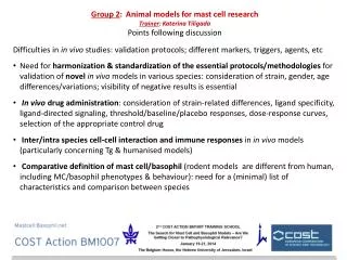
GROUP 5 IN VITRO MODELS FOR HUMAN MAST CELLS. Trainer: Ilan Hammel Trainees: Patricia Valentin Mansour Seaf Adi Efergan Iva Polakovicova Rosa Torres. Introduction. Differences between human and mouse mast cells. Limitation of HuMCs

E N D
GROUP 5IN VITRO MODELS FOR HUMAN MAST CELLS Trainer: Ilan Hammel Trainees: Patricia Valentin Mansour Seaf Adi Efergan Iva Polakovicova Rosa Torres
Introduction • Differences between human and mouse mast cells. • Limitation of HuMCs AIM: To make a comparison between different Human mast cell models MCs Cell lines- HMC-1 LAD-2 Primary cultured MCs- HLMCs sMCs Peripheral derived CD34+ cells ESMCs CBMCs
Introduction • TO BE DISCUSSED: • Similarities vs differences • Gene profile and mRNA expression • Proteins composition and protein levels • Stage of maturation • Granule content • Cell surface receptors expression • Modes of activation and secretion
Vascular endothelial growth factors synthesized by human lung mast cells exert angiogenic effects Aikaterini Detoraki, MD, PhD,a Rosaria I. Staiano, PhD,a Francescopaolo Granata, MD, PhD,a Giorgio Giannattasio, MD,a • Nella Prevete, PhD,a Amato de Paulis, MD,a Domenico Ribatti, MD,b Arturo Genovese, MD,a Massimo Triggiani, MD, • PhD,a and Gianni Marone, MDa
Human mast cells express and synthesize VEGFs • Expression of VEGFs in human mast cells. • VEGF-A, B, C, D and PlGF RT-PCR amplification products from HLMCs, LAD-2 cells, and HMC-1 cells. • Immunoblot with anti–VEGF-B, C, D and anti–GAPDH antibodies of HLMC, LAD-2, and HMC-1 lysates. MCF-7 and RAW 264.7 cells were used as positive controls.
PGE2 and NECA affect expression and release of VEGF in a time-dependent manner HMC-1 LAD-2 HLMCs HLMCs Effects of NECA on VEGF production from human mast cells. A. Expression of VEGF-A, B, C and D was determined in HMC-1 cells by qPCR. B. VEGF-A release was determined in HLMCs by using ELISA. Effects of PGE2 on VEGF production from human mast cells. A. Expression of VEGF-A, B, C and D was determined in LAD-2 cells by qPCR. B. VEGF-A release was determined in HLMCs by using ELISA.
Human mast cells express both VEGFR-1 and VEGFR-2 HLMCs • Expression of VEGFRs in human mast cells. • VEGFR-1, soluble VEGFR-1, VEGFR-2, and GAPDH RT-PCR amplification products from HLMCs, LAD-2 cells, and HMC-1 cells. • The cell surface expression of VEGFRs in HLMCs was determined by FACS.
Human mast cells synthesize and release angiogenin, a member of the ribonuclease A (RNase A) superfamily Marianna Kulka,*,1 Nobuyuki Fukuishi,† and Dean D. Metcalfe†
Human mast cells synthesize angiogenin Analysis of ANG expression. A. HMC, HMC-1, LAD2, and CD34+- derived HuMC expression of ANG and actin by qRT-PCR.
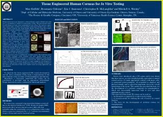
Human embryonic stem cells: a source of mast cells for the study of allergic and inflammatory diseases Martina Kovarova, Anne M. Latour, Kelly D. Chason, Stephen L. Tilley and Beverly H. Koller
ESMCs produce tryptase and chymase in the same manner as CBMCs Phenotype of Embryonic Stem cell-derived mast cells. A. Cells were stained with α- tryptase, α-chymase , and Toluidine blue. B. Quantification of tryptase- and chymase- positive cells in culture during mast cell differentiation. C. Enzymatic activity of tryptase measured in mast cell lysates. D. Inhibition of tryptase activity by tryptase antagonist.
ESMCs and CBMCs express FcεRI and c-Kit on the cell surface • Expression of FcRI and c-Kit on ESMCs. • FACS analysis of ESMCs. • ESMCs differentiated by co-culture with OP9. • B. ESMCs differentiated by EB formation. • C. CBMCs. • D. Expression of c-Kit in ESMCs differentiated by EB formation. • E. Double staining of ESMCs by α-IgE and α-c-Kit antibody. ESMCs- OP9 ESMCs- EB CBMCs
ESMCs with the same genetic modification can be generated Human mast cells differentiated from genetically modified hES cells. Mast cells derived from GFP-hES express tryptase. Top panel: GFP-hES cells. Bottom panels: Mast cells derived from non-transfected H1 clone.
Mast cell lines HMC-1 and LAD2 in comparison with mature human skin mast cells * * Histamine content OK
Cultured Mast Cells from Patients with Asthma and Controls Respond with Similar Sensitivity to IgE-Mediated Activation Table 2 Mast cell numbers obtained from asthmatics and control persons (median and range). • The CD63 MFI and the % CD63+ mast cells cultured from patients with asthma and controls were similar. • The maximal CD63 MFI • The median % CD63+ mast cells • measured on allergen-specific activated mast cells from patients with asthma and controls.
Gene Expression Profiling of Human Mast Cell Subtypes: An In Silico Study Gene Expression Profiles of 16 Samples 2 basophil samples, 2 eosinophil samples, 2 neutrophil samples, 3 lung-derived MC samples, 3 tonsil-derived MC sample, 3 peripheral blood progenitor-derived MC samples, 1 cord blood-derived MC sample
Summary • What are the biological questions that are best addressed by the model that you discussed? • To analyze the function and development of human mast cells. • Mast cells play a pivotal role in bronchial asthma, allergic diseases and are also implicated in other chronic inflammatory conditions and tumor growth. Therefore, human MCs are the most appropriate model for the investigation of human MCs related disease.
Summary • If more than one model exist- do the distinct models yield similar results? • If not, point out the discrepancies. Criticism of the model discussed. • Human mast cells can be derived from isolated CD34+ or CD133+ hematopoietic precursors from either cord blood or peripheral blood – need to be cultured several weeks • Alternatively, mature human mast cells can be isolated from a few human tissues, including lung, skin and gut – can be used immediately • Limitations: • 1. Low number of cells • 2. Cannot be cultured indefinitely- continuous source is required • 3. Genetic differences are present between each population • 4. Primary mast cells cannot be easily genetically manipulated • 5. Long culturing in the same condition affects the fenotype • Suggestions: • Differentiating cells by using co-culture on supporting cell types such as fibroblast feeder layer to provide beneficial microenvironment for the differentiation and proliferation • Human Embryonic stem cell derived MCs offer an alternative– e.g. to follow mast cell differentiation and tracking at in vitro and in vivoexperiments
Summary • If more than one model exist- do you distinct models yield similar results? • If not, point out the discrepancies. • Criticism of the model discussed. • Also, there are 2 human MC lines: HMC-1 and LAD2 • Limitations: • cell-lines • immature MCs • low amount of tryptase and chymase • HMC-1 don’t express FceRI
Summary • Nothing is perfect in life • None of the model is suitable • The choice of the model depends on the interest of your study THANK YOU!!