Download
1 / 26
290 likes | 552 Views
BASIC HISTOLOGY OF THE ENDOCRINE GLAND. DR PAUL. Endocrine System. Introduction . organs synthesize and release chemical messengers – hormones to maintain homeostasis release into the interstitial fluid to enter into the blood stream

E N D
Endocrine System Introduction • organs synthesize and release chemical messengers – hormonesto maintain homeostasis • release into the interstitial fluid to enter into the blood stream • to act on specific target organ (receptor mediated) or cause generalised response • slower, prolonged effect • under the control of nervous system • controls nervous system function and immune system
Types of Hormones • Over 50 hormones secreted • three classes based on the chemical nature • steriod – lipid derived hormones ; e.g. ovarian, testicular and adrenal hormones • small peptides, proteins & glycoproteins - most of the hormones secreted by pituitary, thyroid, parathyroid, pancreas and enteroendocrine cells • - amines e.g. epinephrine and non-epinephrine
Parts of Hypophysis Adenohypophysis (anterior pituitary) - pars distalis, pars tuberalis and pars intermedia Neurohypophysis (posterior pituitary) - pars nervosa, pars infundibularis and median eminence
Histology of adenohypophysis • - remnant of Rathke’s pouch • - rudiment in human • cords of basophilic cells that resemble corticotrophs • synthesize ACTH and MSH, morphin related compoundslike endorphins and lipotrophins • cystic spaces – remnant of Rathke’s pouch
Pars distalis …………………… • two major types of cells based on affinity to stain • chromophobes • chromophils
Chromophobes do not stain intensely - two types of cells : - cells with few secretory granules – resting chromophils / degranulated chromophils - cells with no granules : undiffernetiated cells & follicular cells that have processes for supportive function
Chromophils ……. Acidophils – orange Basophils – blue cells are not evenly distributed; particular cell type is localised in particular part of the gland
Regulation of Hypophysis function 1). Hypothalamic releasing and inhibitory factors releasing factors : TRF , SRF (GRF), GnRF, CRF, PRF inhibitory factors: somatostatin & PIF 2) negative feed back from the circulating hormone e.g. TRF promotes– TSH-promotes---Thyroxin Thyroxin –inhibits secretion of TSH & TRF
Parathyroid • two pairs of gland in the posterior surface of thyroid gland septum parathyroid thyroid
Gross - histology • loose connective tissue fibrous capsule • septa dividing the gland into lobules; carry blood vessels, lymphatics and nerve fibres
Thyroid Follicle • structural and functional unit • spherical, surrounded by reticular fibres with extensive capillary network • lined with simple cuboidal epithelium • Contain gelatinous glycoprotein (thyroid colloid) -thyroglobulin
Structural Variability • inactive gland • - squamous epithelium • - large follicle • -active follicle • - cuboidal or columnar epithelium • - smaller follilcle
Structure of follicular cell • cytoplasm rich in cellular organelles for synthesis and secrete thyroglobulin & thyroid hormone • -cells have microvilli • basal cell membrane has receptors for thyrotrophin
Histological feature Brain sands Neuroglial cells pinealocytes Parenchyma has typical endocrine form of organisation - cells are arranged in the form of cords surrounded by fenestrated capillaries
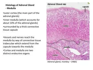
Adrenal Gland Supra Renal Gland Location : superior pole of kidney Parts: cortex and medulla Stroma: reticular fibres outer cortex medulla capsule
Steroid secreting portion of the gland; maintains ion and water homeostasis; carbohydrate, protein and lipid metabolism Cortex Capsule Three zones: Zona glomerulosa Zona fasciculata Zona reticularis Medulla
Zona Glomerulosa • ovoid cluster of cells with delicate trabeculae inbetween • columnar or pyramidal cells • acidophilic cytoplasm • few lipid droplets • secrete mineralocorticoids - aldosterone ZG Function: increases sodium and water absorption by kidney tubules to maintain ion and fluid homeostasis ; to increase blood pressure - act on GI mucosa, salivary ducts, sweat gland to increase sodium absorption Secretion is controlled by renin-angiostensin and NOT by ACTH
Zona Fasciculata • polyhedral cells arranged as radiating cords of 1 or 2 cells thick • - fenestrated capillaries between the cords • large round nucleus • pale staining cytoplasm because of abundant lipid droplets • secrete glucocorticoids (cortisol- hydrocortisone and corticosterone) • small amount of androgen – does not cause any appreciable effect on testis
Zona Reticularis • branching cords of cells forming irregular network with wide diameter capillaries in between them • cells are smaller with scanty lipid droplets in the cytoplasm • secrete small quantities of androgen (dehydroepiandrosterone& androstenedione) and glucocorticoids • lipofuscin pigments – age pigments (degradation product of cellular organelle) are notable feature
Adrenal Medulla • - polyhedral cells in clusters • cells are also called chromaffin cells as the secretory granules are oxidised to give brown color in fixation with chrome salts. • basophilic granular cytoplasm • scanty endoplasmic reticulum • no stored lipids • stroma – fine collagen fibres • abundant sinusoidal capillaries
Two types of cells • N : norephinephrin secreting cells; dense core, large granules. • A: epinephrine secreting cells ; smaller less electron dense granules; outnumber N cells in population • Norepinephrin + N-methyl = epinephrin • cells also secrete enkephalin – endogenous opiate • chromogranin (with ATP and Ca2+) binds low molecular weight catecholamines • dopamin beta hydrolase (for dopamin conversion into norepinephrin
“Fight- Flight response” Organism’s defense reaction against threat physiological and psychological stress (unprepared exam fever) Cause: sudden spurt of catecholamine release- establish a condition for maximum use of energy to generate maximum physical effort ( to fight or run away) Effect: stimulate glycogenalisis and mobilisation of free fatty acid from adipose tissue Overt reaction: increased heart rate and cardiac output, dilatation of coronary artery, bronchiole dilatation, increases respiratory rate, vasodilatation of vessels to skeletal muscle, vasoconstriction of blood vessels to smooth muscle of gut and skin, increased sweat activity
Adrenal disorders • adrenal medulla tumor: pheochromocytoma- hyperglycemia and transient hypertension • adrenal cortex disorder: - hypertrophy OR • hypotrophy • Hypertrophy (excessive production of steroid)- Cushing syndrome or Conn syndrome; secondary sexual disorders • Hypotrophy: autoimmune disease (insufficient secretion of steroids) - Addison disease
Islets of Langerhans A cells: regular granules; peripherally located; secrete glucagon- elevate blood sugar level by stimulating glycogenolisis B cells: irregular granules; centrally located ; secrete insulin – promote uptake of glucose by liver cells, skeletal muscle and adipose cells to decrease blood sugar level D cells: somatostatin secreting cells