Download

1 / 42
550 likes | 1.73k Views
Primary Dentition. Dental Formulas. Shorthand summary of teeth present Maxillary quadrant/mandibular quadrant Multiply by 2 for entire dentition. Examples of Dental Formulas. Human permanent dentition I 2/2 C 1/1 P 2/2 M 3/3 Human primary dentition I 2/2 C 1/1 M 2/2 Cats

E N D
Dental Formulas • Shorthand summary of teeth present • Maxillary quadrant/mandibular quadrant • Multiply by 2 for entire dentition
Examples of Dental Formulas • Human permanent dentition I 2/2 C 1/1 P 2/2 M 3/3 • Human primary dentition I 2/2 C 1/1 M 2/2 • Cats I 3/3 C 1/1 P 3/2 M 1/1 • Dogs I 3/3 C 1/1 P 4/4 M 2/3
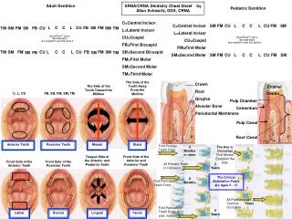
Numbering of Primary Teeth • Universal system: A to T • Palmer: A to E by quadrant • FDI: 1st number 5– 8, second 1 to 5
Dentition Periods • Primary dentition period: only primary teeth are present, 6 months to 6 years • Mixed dentition period: the primary teeth are being replaced by the permanent teeth, 6 to 12 years • Permanent dentition period: after the last primary tooth has exfoliated
Dentition Periods • Primary • Mixed • Permanent
Clinical question: Why is it important to maintain the health of the primary teeth? • Mastication of solid foods • Speech development • Esthetics and self-esteem • Space for eruption of permanent teeth • Health of the permanent teeth
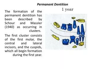
Eruption of Permanent Teeth • 1st molars are first permanent teeth to erupt. • Referred to as 6 year molars • They erupt distal to primary dentition.
Eruption Sequence • In general, mandibular teeth erupt before maxillary counterpart. • 1st molars are first permanent teeth to erupt. • Central incisors are first SUCCEDANEOUS teeth to erupt. • Note the late eruption of the maxillary canine: may be impacted due to space loss. Succedaneous teeth are permanent teeth that replace primary teeth. AB D C E A B D CE
General Differences Between Primary and Permanent Teeth • Size: smaller in all dimensions. • Color: lighter in color. • Crowns: bulbous, wider mesiodistally, shorter incisocervically • CEJ: appears constricted • Roots: longer and more slender
Primary – Permanent Comparisons: Differences in Internal Anatomy Primary teeth have: • thinner enamel & dentin • relatively larger pulp cavities Pulp horns are closer to outer surface Great variation in size & location Form of pulp follows external anatomy Usually a pulp horn under each cusp **Mesial pulp horn is higher
Primary Posterior Teeth • Short, bulbous crowns • Slender, flared roots • Short root trunk • Second molars > first molars
Maxillary First Molar(Deciduous) GROUP 6: Celso, Anne R. Flores, Joyce Anne G. Jung, Young Min Parmar, Asma Ramirez, Jan Andre P.
Primary Maxillary Molars • 3 roots • 1st resembles a maxillary premolar • 2nd resembles a permanent maxillary 1st molar
Buccal Aspect • Mesial half of the crown has a greater height • DBR,MBR, and palatal root • Slender and long • Spread widely
Buccal Aspect • Distal root is shorter than Mesial root • Lingual root is positioned midway bet. the Bu roots
Buccal Aspect • Bifurcation of the roots begins almost immediately at the Cervical line • Little root trunk
Lingual Aspect • Almost similar to buccal aspect • MLC is prominent • DLC is poorly defined • DBC visible
Lingual Aspect • All roots visible • Li root is larger MD • Li surface is entirely made up one cusp • Slightly convex occlusocervically, but markedly convex MD
Occlusal Aspect • MB, DB line angle are greater than ML,DL line angle • Mesial line angles are greater than Distal line angles • Crown converges lingually and distally • Occlusal surface is nearly rectangular • Has a center fossa and pit
Occlusal Aspect • Has BG and LG • Has a Mesial Triangular Fossa • Mesial pit • No distal pit • Supplementary groove in the mesial pit
Mesial Aspect • Cervical width is much greater than occlusal due to a very promi- nent cervical ridge • Buccal outline is straight or slightly concave
Mesial Aspect • Lingual outline is convex • ML cusp is longer and sharper than MB cusp • Cervical line is slightly curved toward occlusal
Mesial Aspect • Marginal ride is shorter and less prominent
Distal Aspect • Smaller than mesial aspect • MR is less prominent • Db cusp is longer and sharper than DL cusp
Distal Aspect • Cervical line straight and slightly curved occlusally • Cervical ridge is not so prominent
Primary Mandibular Molars • 2 roots • 2nd resembles the mandibular 1st permanent molar • 1st does not resemble any other permanent or primary tooth!
Summary • Primary teeth have • Thinner enamel and dentin layers • Pulp horns closer to the outer surface • Mesial pulp horn much higher • Relatively large pulps • Enamel rods directed slightly occlusal in cervical area • More tortuous and irregular pulp canals