Download
1 / 22
230 likes | 551 Views
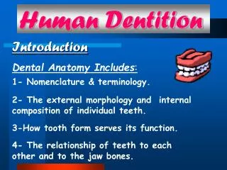
BIOLOGY OF THE HUMAN DENTITION. Temporomandibular Joint -TMJ. Anatomy of TMJ. Ligaments: There are three ligaments associated with the TMJ:one major and two minor ligaments.

E N D
BIOLOGY OF THE HUMAN DENTITION Temporomandibular Joint -TMJ
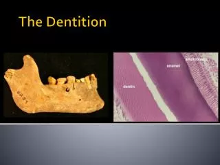
Anatomy of TMJ Ligaments: There are three ligaments associated with the TMJ:one major and two minor ligaments. The major ligament, the temporomandibular ligament, or lateral ligament, is really the thickenest lateral portion of the capsule. Some authors mention the temporomandibular ligament as an anterior thickening of the capsule, not a separate ligament.
Anatomy of TMJ capsule lateral ligament
Anatomy of TMJ The minor ligaments, • the stylomandibular ligament, This ligament runs from the styloid process to the angle of the mandible. stylomandibular ligament
Anatomy of TMJ • the sphenomandibular ligament. It arises from the angular spine of the sphenoid bone and ends broadly at the lingula of the mandible. sphenomandibular ligament
Anatomy of TMJ Both minor ligaments are accessory and are not directly attached to any part of the joint. They may stabilize the articular system during jaw movements. These ligaments are important for defining the border movements, or in other words, the farthest extents of movements, of the mandible.
Anatomy of TMJ However, movements of the mandible made past the extents functionally allowed by the muscular attachments will result in painful stimuli.
Anatomy of TMJ Muscles: The mandible is moved primary by the four muscles of mastication: • the masseter, • the medial pterygoid, • the temporalis and • the lateral pterygoid.
Anatomy of TMJ Lateral pterygoid muscle Medial pterygoid muscle Masseter muscle
Anatomy of TMJ Temporalis muscle Lateral pterygoid muscle Masseter muscle
Anatomy of TMJ These four muscles, all innervated by the mandibular division of the trigeminal nerve, work in different groups to move the mandible in different directions.
Temporalis muscle • Origin: temporal fossa & temporal fascia • Insertion: coronoid process & anterior of ramus • Function: Elavation and positioning of the mandible Temporalis muscle
Masseter muscle • Origin: zygomatic arch • Insertion: lateral surface of ramus, coronoid process & angle of mandible • Function: power with vertical elevation of the mandible; deep portion stabilizes the condyle in protrusive closure
lateral pterygoid muscle • Function of the inferior portion: protrusion, lateral movement and contributes to opening • Function of the superior portion: active with themuscles of closure, especially aiding stabilization of the condyle during the power stroke.
Medial pterygoid muscle • Function: elevation of the mandible, protrusion of the mandible and lateral movement of the mandible with unilateral activation
Anatomy of TMJ Contraction of the lateral pterygoid acts to pull the disc and condyle forward within the glenoid fossa and down the articular eminence; thus, action of this muscle serves to open the mouth.
Motion of TMJ The TMJ is an example of diarthrosis, and its movements are a combination of: • a hinge action and • a gliding action.
Motion of TMJ The lower joint compartment formed by the mandible and the articular disk is involved in rotational/hinge movement (opening and closing movements). For the first 15° to 18° of motion, the condyle moves without disk movement. The condyle simply rotates in the capsule; a purely hinge motion.
Motion of TMJ With a motion past 18°, the disk and condyle begin moving forward and down the articular eminence of the temporal bone. The condyle is now rotating and gliding down the articular eminence; a hinge and gliding joint. The upper joint compartment formed by the articular disk and the temporal bone is involved in this translational movements.
Motion of TMJ At maximum opening, the condyle and disk are positioned inferior to their original position, the condyle has rotated such that the posterior portion of the head is now articulating with the temporal bone, and the medial and lateral pole attachments of the disk have dragged it down with the condyle while the retrodiscal lamina fibers have stopped the disk from moving further over the condyle.