Download
1 / 60
630 likes | 815 Views
The Cell Cycle Jim Umen BGGN222 Feb. 21, 2006. Outline of Class. 1. History and fundamentals. 2. MPF and the discovery of CDKs. 3. CDK regulation of mitotic entry and exit. 4. Regulation of G1-S. 5. DNA replication control. 6. Cell cycle checkpoints. 7. Discussion of papers.

E N D
Outline of Class 1. History and fundamentals 2. MPF and the discovery of CDKs 3. CDK regulation of mitotic entry and exit 4. Regulation of G1-S 5. DNA replication control 6. Cell cycle checkpoints 7. Discussion of papers
Key points 1. Essential components of the cell cycle and what they do 2. Logic of cell cycle circuitry and switches 3. Experimental approaches Many topics we have no time to cover, or will cover in little detail: chromosome dynamics, cytokinesis, centriole replication, cancer cell cycles, developmental/alternative cell cycles, checkpoints, DNA replication mechanisms, meiotic cell cycle, growth control . . . .
What is a Cell Cycle? Process by which cells replicate themselves 1830’s Schleiden & Schwann organisms are made of cells 1850’s Schultze cytosol (protoplasm) and nucleus defined as separate entities in animal and plant cells 1850’s Remak and Virchow omnis cellula e cellula --all cells come from other cells 1880’s Fleming and Strasburger mitosis (chromosomes look like threads) 1950’s Stages of cell cycle are defined DNA replication as a discrete event (S phase) 1960’s Continuous and discontinuous events RNA synthesis, protein synthesis, cell growth--continuous DNA synthesis, Mitosis--discontinuous
Protein Mitosis RNA DNA Mass/Size 2 M Relative Amount 1 G2 G1 G1 S G2 M Restriction Point time S Interphase Cell Cycle Fundamentals 1 S phase and Mitosis are defined by processes G1 and G2 (gap phases) are defined by timing G1 (and G2?) can be split into more meaningful sub-stages by molecular and physiological criteria: Restriction Point in mammalian tissue culture cells defined by serum sensitivity
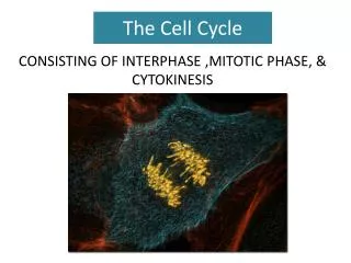
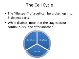
Cell Cycle Fundamentals 2 Mitosis is subdivided into different stages
1. DNA replication S phase S phase 2. Centrosome duplication (microtubule organizing center) 3. Nuclear Division (karyokinesis) Mitosis Mitosis 4. Cell Division (cytokinesis) 5. Cell Growth Throughout Cell Cycle Fundamentals 3 The cell cycle can be broken into subcycles whose relationships change in different cell types and under different circumstances Timing in a typical somatic cell: In a typical somatic cell 1-5 are all regulated and coupled
1. DNA replication 2. Centrosome duplication (microtubule organizing center) 3. Nuclear Division (karyokinesis) 4. Cell Division (cytokinesis) 5. Cell Growth Cell Cycle Fundamentals 4 Non-canonical cell cycles are found throughout nature and play a critical role in cell and developmental biology Somatic cell 1-5 regulated Embryonic cell cycle: 1,2,3,4 (division with no growth) Meiotic cell cycle 2,3,4: (division with no DNA replication) Megakaryocytes, slime molds: 1,2,3,5 (replication and nuclear division) Liver cells, fly salivary glands, many plant tissues: 1,2,5 (endoreplication=S phase with no mitosis) Oocyte formation: 5 (growth with no division) Some green algae: 5 and 1,2,3,4 separated (Chlamydomonas) or 1,2,3,5 and 4 separated (Scenedesmus) Ciliated epithelial cells, some protozoans: 2 (centriole amplification)
Cell Cycle Fundamentals 5 Cell Cycle States are Regulated I. Johnson and Rao (1975) Cell fusion experiments
Starved plasmodium no growth, no DNA replication UV irradiate ~50% of nuclei inactivated Remaining nuclei replicate and divide until nucleo-cytoplasmic ratio is restored Cell Cycle Fundamentals 5 Cell Cycle States are Regulated: nucleocytoplasmic ratio controls replication II. (Sudbery and Grant, 1976) Physarum (slime mold)
Cell Cycle Fundamentals 6 Models for Cell Cycle Analysis Xenopus oocytes/extracts Simple biochemical system budding yeast (Saccharomyces cerevisiae) Powerful genetics, G1-S regulation fission yeast (Schizosaccharomyces pombe) Powerful genetics, G2-M regulation mammalian tissue culture cells e.g. HeLa cells, NIH3T3 Closest model for human cells, regulation is more complex Details vary between organisms, general principles are similar
Xenopus oocytes/extracts Properties of Xenopus system: Cell free extracts can cycle between S phase and Mitosis Extracts can be manipulated to effect a mitotic arrest: chelate calcium > stabilize cytostatic factor (CSF) Feedback controls/checkpoints are missing* -extracts can cycle without nuclei G1 regulation is absent
MPF activity fluctuates and is present in both oocytes and fertilized embryos Cyclin synthesis and abrubt degradation mirrors rise and fall of MPF activity Tim Hunt and co-workers Discovery of MPF (maturation promoting factor) Serial injections can be repeated indefinitely until original source of MPF is diluted away Yoshio Masui and co-workers
Lohka and Maller Stabilize MPF in vitro from Xenopus extracts and fractionate: MPF has two components 32 kd and 45 kd; MPF posseses kinase activity 45 kd protein = cyclin B Kinase activity only present when both subunits are present Take cell free extract and RNase treat to destroy all endogenous mRNAs Murray and Kirschner Inactivate RNase with inhibitor and add no RNA or cyclin mRNA activate extract to induce interphase and add sperm nuclei Biochemical nature of MPF I Several groups:
∆13 equiv. to wt cyclin B ∆90=nondegradable cyclin missing destruction box Murray and Kirschner cyclin MPF Biochemical nature of MPF II Cyclin synthesis drives activation of MPF Cyclin destruction is required to inactivate MPF and drive cells into interphase MPF is required for cyclin destruction Oscillator behavior of cell cycle is explained: Cyclin destruction is NOT required to initiate anaphase
Yeast Genetics and Unification of the Cell Cycle Main control point is G2/M boundary Main control point is G1-S boundary
cdc28 (budding yeast) has two arrest points pre-S phase, and pre-M cdc2 (fission yeast) shows arrest at G2/M, but also has dominant alleles that give a wee phenotype phenotypes suggest that these two cdcs have a critical role in cell cycle regulation cdc mutants are critical for identifying cell cycle components Hartwell and colleagues, Nurse and colleagues budding yeast fission yeast G1-S blocked cdc mutant mitotic cdc mutant
Unifying observations for the cell cycle field budding yeast CDC28 = fission yeast cdc2 = Xenopus MPF 32kd subunit aka CDK1 budding yeast daf1/whi1 = a cyclin (later renamed Cln3) fission yeast cdc13 = a B-type cyclin homolog Key enzyme for cell cycle regulation is now defined as a cyclin dependent kinase complex (CDK) composed of a catalytic kinase subunit and a cyclin that activates the kinase Since then things got more complicated: Multiple CDKs, Multiple cyclins and Interacting proteins discovered All eukaryotes use the same set of proteins for cell cycle regulation with some species specific variation 2001 Nobel Prize in Physiology or Medicine given to Tim Hunt, Lee Hartwell, and Paul Nurse for their pioneering work in cell cycle regulation
Understanding the somatic cell cycle Somatic cells and yeasts have a G1 period with low CDK activity Somatic cells and yeasts have multiple cyclins or CDKs that control progression through G1, S phase and mitosis Somatic cells and yeasts have feedback controls that gate each transition to ensure proper completion of previous events
CDK Regulation How is CDK activity controlled during the cell cycle? Activation by cyclin binding Activation by phosphorylation (CAK) Activation by dephosphorylation (CDC25, Cdi1) Activation by destruction of inhibitor (Skp1-Cullin-F box complex aka SCF) Inactivation by cyclin destruction (Anaphase Promoting Complex/Cyclosome aka APC/C) Inactivation by phosphorylation (Wee1) Inactivation by inhibitory binding proteins (KIP/CIP/WAF/KRP and INK) Substrate specificity (CDK-cyclin combinations) Subcellular localization CDK abundance usually not regulated
CDK regulation I Why is CDK kinase activity non-linear with respect to cyclin concentration? Cyclin concentration or CDKactivity time
Inhibitory kinase Activating phosphatase wee1 cdc25 Cell cycle target proteins CDK activation and inactivation by feedback loops CAK (CDK Activating Kinase) CKI/KIP ICK/KRP Cdc2-cyclin B Ubiquitin ligase for cyclin APC/C
Inactive monomer ATP misoriented, substrate binding occluded by T-loop, PSTAIRE helix mispositioned Cyclin bound ATP properly oriented via interaction with repositioned T loop and PSTAIRE helix. Substrate binding cleft suboptimal. Tyr14 site in roof of ATP binding cleft is available for Wee1phosphorylation (not shown) Structural basis for CDK activation by cyclins and phosphorylation I
CDK Thr160 +cyclin T loop flattened. Phospho T160 forms stabilizing interactions that optimize binding site With substrate peptide SPXK-containing peptide fits into pocket and interacts with T loop, including Phospho T160. Structural basis for CDK activation by cyclins and phosphorylation II
CAK (CDK activating kinase) --largely unregulated Wee1 Major negative regulator Cdc25 Major positive regulator Balance between Cdc25 and Wee1 activities regulates mitotic entry Phosphorylation/Dephosphorylation of CDKs cdc2
active inactive inactive active active inactive Postive Feedback Loop for CDK Activation Loop leads to explosive auto-activation of CDK once its activity rises above a certain threshold What are the substrates CDK1-cyclin B that lead to mitotic entry and progression? Cdc25, Histone H1, lamins, cyclin B and many more
KEN RXXL Mitotic Exit is Regulated APC/C I APC/C is a E3 specificity factor for ubiquitin ligase pathway examples: B cyclin Cdc20 Cdc20 + + or Pds1 or Cin8 Hct1 KEN-box substrates Hct1 only activator and specificity factor RXXL destruction (D) box- containing substrates Cdc20, Hct1 (Cdh1) core complex Targeting by APC/C leads to rapid degradation by the 20S proteasome
Mitotic Exit is Regulated APC/C II B Cyclins and Pds1(Securin) are key substrates of APC/C: Nondegradable cyclin blocks MPF destruction but does not block anaphase APC/C has at least one more target whose destruction promotes anaphase Pds1/Securin destruction releases a protease, separase, that degrades cohesisns and allows sister chromatids to separate
Budding yeast mitotic exit Decreased CDK activity and Separase release activate FEAR (Cdc14 early anaphased release) Cdc14 is a protein phosphatase that plays a central role in exiting mitosis Cdc14 dephosphorylates and activates cdh1 subunit of APC and Sic1 (a CDK inhibitory protein) to establish a stable G1 state with low CDK activity Cdc14 activates a second pathway called MEN (mitotic exit network) that initiates cytokinesis
Overview of APC activation and mitotic exit CDK1-Cyclin B activates APC-Cdc20 directly or indirectly through phosphorylation A time lag between APC-Cdc20 activity and other essential mitotic events is essential Decreased CDK activity allows activation of Cdc14 mitotic exit pathway, Cdh1/Hct1 and establishment of a stable G1 state Regulation of APC/C by CDK1-Cyclin B generates a negative feedback to drive mitotic exit
G1 and G1-S regulation G1 is characterized by low CDK activity and high APC-Cdh1 activity What triggers initiation of S phase and cell cycle re-entry?
G1 and G1-S regulation I G1 is a major control point for most cell types: Growth factors present and extracellular conditions favorable: S phase G1 M Differentiation factors present, unfavorable conditions: G0 (temporary or permanent withdrawal from cell cycle) What triggers entry to S phase, what mechanisms prevent it? Cells must be growing and have reached a minimum size mammalian cells must not contact neighbors (contact inhibition) APC-Cdh1 must be inactivated to allow S phase cyclin accumulation CDK inhibitory proteins must be destroyed or titrated away In budding yeast and animal cells G1 CDK activity must reach a threshold value to trigger S phase
Pardee (1974) Serum Dependent Serum Independent Restriction point Restriction point in animal cells occurs late in G1 S G1 Serum withdrawal before R point--cells arrest in G1 Serum withdrawal after R point-cells complete S, G2 and M Budding yeast cells have a G1 control point termed Start Remove nutrients prior to Start--G1 arrest Remove nutrients after Start--cells complete S, G2 and M Start G1 control points
animal cells budding yeast Growth factors in serum e.g. FGF,PDGF nutrients (glucose, nitrogen etc.) increased protein translation rate activation of RTK signaling Increased translation of Cln3 transcription of D cyclins G1 and G1-S regulation II Similarities of Cln3 and D cyclins: messages and proteins are low abundance proteins are highly unstable length of G1 highly sensitive to dosage and expression levels control rate limiting step in G1-S transition neither are essential!
Cullin Skp1 Cdc4 is F box adaptor for Sic1 F-box protein (adaptor) substrate Triggering Start in budding yeast I G1 cyclins Cln1, Cln2, Cln3 Sic1--CDK inhibitor--disrupts CDK active site, prevents ATP binding Transcription factors SBF, MBF--activators of Cln1, Cln2 and other S phase genes Whi5--negative regulator of SBF, MBF SCF--Skp1-Cullin-Fbox--E3 ubiquitin ligase targets G1 substrates (Elledge and Harper 1996) F-box proteins are specificity factors in SCF, often require phosphorylation for binding target In early G1: Sic1 and APC-Hct1/Cdh1 are dephos. and active. CDK activity is low
As Cln3-Cdc28 activity builds: Whi5 is phosphorylated and dissociates from SBF/MBF SBF/MBF become active Cln1/2 and Clb5/6 are made SBF/MBF are further activated in a positive feed back loop Sic1 and Hct1 are inactivated in a negative feedback loop Triggering Start in budding yeast II Cln3-Cdc28 Whi5 SBF or MBF Cln1 Cln2 ? Clb5 Clb6 Cln1/2-Cdc28 SCF-Cdc4 Sic1 Clb5/6-Cdc28 Hct1/Cdh1
Cdc4 binding of Sic1 depends on 6+ phosphorylations Replacement with a high affinity Cdc4 binding site causes premature S phase initiation and genome instability Tyers and colleagues Sic1 inactivation is key for S phase initation Triple mutant ∆cln1 cln2 cln3 is inviable ∆cln1 cln2 cln3 sic1 mutant--viability is rescued! Sic1 is key target of G1 cyclins Sic1 becomes multiply phosphorylated by CDKs during G1 followed by abrupt degradation
G1 control in mammalian cells G1 cyclins D1-D3-(like Cln3) CDK4, CDK6-specific for D cyclins G1-S cyclin E (like Cln1/2) CDK2-binds E and A cyclins CDK inhibitors p27 Kip1 (homologous to Sic1), INK family (no homolog in yeast) E2F complexes (transcription factors for S phase genes, Cyclin E (like SBF/MBF) RB/p107/p130--E2F repressors (like Whi5) SCF-Skp2--targets free cyclin E and p27 for degradation (like SCF-Cdc4)
Establishing functions of G1 CDKs and cyclins Can’t easily make cdc mutants with diploid mammalian cells Strategies for genetic analysis: dominant negatives overexpression (transfection) knockouts in ES cells, whole mice, or cell lines from KO mice siRNA-mediated knockdowns Harlow and colleagues Dominant negative CDK mutations For many years CDK2/CycE thought to be a linchpin of G1-S regulation However, CDK2 and CycE KO cells have only mild S phase entry defects!
Negative regulation of G1-S is critical for animal cells Unregulated cell division leads to defects in tissue morphogenesis, development and cancer Several G1 regulators are tumor suppressors or oncogenes-RB, INKs, D cyclins, CDK4 Two classes of negative regulators: CDK inhibitors INK4 (CDKN2) family specific for CDK4/6-Cyclin D complexes p21, p27,p57 proteins inhibit all CDKs RB-related proteins RB, p107, p130--bind to E2F complexes and repress S phase transcription Regulators show some functional overlap and tissue specificity e.g. RB is expressed in cycling cells, p107/p130 in quiescent cells, p27 is constitutively expressed,p21 is induced by checkpoint activation, p57 is expressed in neuronal cells, p16INK4b induced by negative growth factor TGF beta
CDK CDK cyclin cyclin Conformation change in CDK blocks cyclin binding Binds CDK-cyclin, blocks ATP binding and substrate access Kip INK INKs and KIPs inhibit CDKs in different ways Early G1--INKs keep CDK4/6 cyclin D inactive, p27 keeps CycE-CDK2 inactive As CycD accumulates it overcomes INK binding to CDK4/6 CycD-CDK4/6 complexes compete p27 from CycE-CDK2 promoting S phase
CycE RB P P P P P P P P P P CycA E2F-DP S phase genes RB RB E2F-DP RB E2F-DP E2F-DP Mid G1 RB partially phosphorylated by CDK4/6-D cyclins (priming) Restriction point/late G1/S RB hyperphosphorylated by CDK2-CyclinE complexes dissociation from E2F-DP Early G1 RB hypophosphorylated Phosphorylation of RB is a key step in S phase activation Loss of one copy of RB leads to tumors Animal DNA tumor viruses produce proteins (e.g. SV40 T antigen) that inactivate RB Plant DNA viruses have evolved the same trick CycD and CycE overexpressed in many cancers
Time lapse videomicroscopy on single cells + immunofluorescence to look at timing of RB phosphorylation vs. R vs. S phase Zetterberg and colleagues Does RB phosphorylation=Restriction Point? Previous work on bulk synchronized cells indicates correlation between RB phosphorylation, Cyclin E transcription and Restriction Point What is the molecular correlate of the Restriction point?
G1 control in mammalian cells CycA-CDK2 also blocks E2F DNA binding by phosphorylation
Parallel Mechanisms of G1 regulationin budding yeast and metazoans
Coupling cell size to cell cycle progression yeasts show evidence of size control nutritional shift experiment: move cells from rapid growth to slow growth conditions- observe a G1 (budding yeast) or G2 (fission yeast ) delay until a minmal size is reached Data on animal cells is controversial but evidence for G1 size control exists. Growth may also be cell cycle regulated: RB controls rRNA synthesis Tumors often have aberrant growth characteristics faster cell cycle, faster growth
Regulation of DNA replication I Properties of DNA replication in eukaryotes: Occurs at a specific phase of the cell cycle--S Initiates from specific locations termed origins--well defined in budding yeast poorly defined in other organisms Occurs once and only once per S phase Completion of S phase is ensured by checkpoints S phase is regulated by oscillating CDK activity Low CDK activity required to prime origins High CDK activity required to fire origins and block re-priming
Key Components of S phase Regulation Cdc6 and Cdt1-- origin priming proteins-activity is tightly regulated Orc--origin recognition complex--binds origins throughout cell cycle, required for origin firing Mcms--(mini-chromosome maintenance)-part of a hexameric origin unwinding complex required for initiation, AAA ATPase family Cdc7-Dbf4--kinase complex analogous to CDK-cyclin required for origin firing Geminin (metazoans only) inhibits Cdt1 mediated MCM loading at origins S phase CDKs--Clb 5/6-Cdc28 in budding yeast, CycE-Cdk2, CycA-Cdk2 in mammals
Naked DNA +interphase Xenopus extract Chromatin assembly,NE assembly,1 round of DNA replication Mitosis Next round ofDNA replication Add replicated G2 nuclei to freshinterphase extract +NE permeabilizing detergent lysolecithin control Next round of DNA replication No replication How to ensure one round of replication? Origin “Licensing” Blow, Laskey and coworkers Something present in early interphase extracts that allows replication: licensing factor Licensing factor cannot cross NE. Lf gets made in early interphase, destroyed during S remade during M