Download
1 / 36
360 likes | 505 Views
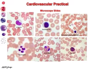
Cardiovascular practical Block. Part I Shaesta Naseem. Normal anatomy and histology . NOTE: The heart serves as a mechanical pump to supply the entire body with blood, both providing nutrients and removing waste products. The great vessels exit the base of the heart.

E N D
Cardiovascular practical Block Part I ShaestaNaseem
NOTE: • The heart serves as a mechanical pump to supply the entire body with blood, both providing nutrients and removing waste products. • The great vessels exit the base of the heart. • Blood flow: body→venacava→right atrium→ right ventricle→lungs→leftatrium→leftventricle→body • The heart consists of 3 layers – the endocardium, myocardium, and epicardium. The epicardium (bottom left) consists of arteries, veins, nerves, connective tissue, and variable amounts of fat. • The myocardium contains branching, striated muscle cells with centrally located nuclei. They are connected by intercalated disks (arrowheads).
Cardiovascular practical Block 1-Atheroma of aorta
These three aortas demonstrate mild, moderate, and severe atherosclerosis from bottom to top. At the bottom, the mild atherosclerosis shows only scattered lipid plaques. The aorta in the middle shows many more larger plaques. The severe atherosclerosis in the aorta at the top shows extensive ulceration in the plaques. • The major risk factors are hyperlipidemia , hypertension , cigarette smoking and diabetes • Complications are thrombosis , hemorrhage , calcifications and aneurysmal dilatation with the distal ischemic events .
This is severe atherosclerosis of the aorta in which the atheromatous plaques have undergone ulceration along with formation of overlying mural thrombus.
Cardiovascular practical Block 2-Coronary atherosclerosis
Coronary atherosclerosis:Cross section of a coronary artery shows: • Partial occlusion of the lumen by an atheromatous plaque. • The plaque consists of cholesterol clefts, hyaline fibrous tissue and some blood capillaries. • The internal elastic lamina is thin and fragmented. • Pressure atrophy of the media opposition atheromatous plaque.
Cardiovascular practical Block 3-Aneurysm of abdominal aorta
Here is an example of an atherosclerotic aneurysm of the aorta in which a large "bulge" appears just above the aortic bifurcation.
Aneurysmal dilatation of lower aorta with rupture , intraluminal thrombus and Extensive aortic atherosclerosis.
The most likely causes of aneurysms are • atherosclerosis • mycotic, • syphilitic and • congenital
Cardiovascular practical Block 4-Myocardial infarction
Complications that might occur: • arrhythmias • ventricular aneurysm • rupture of myocardium, • cardiac tamponade.
Myocardial infarction left ventricles right ventricles Cross section of the left and right ventricles shows a pale and irregular focal fibrosis in the left ventricular wall with increased thickness .
Myocardial infarction:Section of myocardial shows: • Patchy coagulative necrosis of myocardial fibers. The dead muscle fibers are structure less and hyaline with loss of nuclei and striations. • Chronic ischemic fibrous scar replacing dead myocardial fibers . • The remaining myocardial fibers show enlarged nuclei due to ventricular hypertrophy .
Cardiovascular practical Block 5-Left ventricular hypertrophy
Heart from a hypertensive patient. The left ventricle is very thick (over 2 cm). However the rest of the heart is fairly normal in size as is typical for hypertensive heart disease. The hypertension creates a greater pressure load on the heart to induce the hypertrophy
In cross section, this view of the heart shows the left ventricle in the center left of the picture. The heart is from a severe hypertensive. The left ventricle is grossly thickened. The myocardial fibers have undergone hypertrophy.
Heart, normal Heart, left ventricular hypertrophy
Cardiovascular practical Block 6-Vegetations of rheumatic fever on mitral and aortic valves
The small verrucous vegetations seen along the closure line of this mitral valve are associated with acute rheumatic fever. • These warty vegetations average only a few millimeters and form along the line of valve closure over areas of endocardial inflammation. • Such verrucae are too small to cause serious cardiac problems.
Rheumatic valvulitis: Section of fragments of endocardial valve shows: • Irregular endocardial surface, no endocardial lining and focal fibrin deposits. • The valve is thickened by dense hyalinized fibrous tissue with vascularization and chronic inflammatory cell infiltrate
Cardiovascular practical Block 7-Acute rheumatic myocarditis
Acute rheumatic myocarditis:Section of cardiac muscle shows: • Aschoff bodies in the intermuscular fibrous septa. They are oval in shape and seen in relation to blood vessels. • Each consists of a focus of fibrinoid necrosis, few lymphocytes, macrophages and few small giant cells with one or several nuclei (Aschoff giant cell).