Download

1 / 56
600 likes | 1.13k Views
Salivary Gland. Tumors. Classification. Tumors Adenoma (Benign) Carcinomas (malignant) Others. Adenoma (Benign). Pelomorphic adenoma Warthin tumor Basal cell adenoma Oncocytoma Ductal papilomas Canalicular adenoma Sabaceous adenoma Cyst adenoma. Carcinoma (malignant).

E N D
Salivary Gland Tumors
Classification Tumors Adenoma (Benign) Carcinomas (malignant) Others
Adenoma (Benign) • Pelomorphic adenoma • Warthin tumor • Basal cell adenoma • Oncocytoma • Ductal papilomas • Canalicular adenoma • Sabaceous adenoma • Cyst adenoma
Carcinoma (malignant) • Mucoepidermoid ca • Acinic cell ca • Adenoid cystic ca • Ca arising in Pelomorphic adenoma • Polymorphous low grade adenoca • Basal cell carcinoma • Sebaceous carcinoma • Cystadenocarcinoma
others • Soft tissue tumors Haemangioma • Lymphoma Hodgkin lymphoma Diffuse large B-cell lymphoma Extranodal marginal B –cell lymphoma
General Features • Diverse histopathology • 1% of all the tumors of human body • 2% to 5% of head and neck neoplasms
Distribution of Tumors • Parotid: 78% overall; (85%) 75% benign Pleomorphic adenoma 10% other benign 15% Malignant
Distribution of Tumors • Submandibular: 12% overall; 30% Malignant
Distribution of Tumors • Sublingual: 0.3% overall; 14% benign 86% Malignant
Distribution of Tumors • Minor S Gland: 10% Overall; 55% Benign 45% Malignant
Pelomorphic adenoma • Most common of all salivary gland neoplasms • 70-80% of parotid tumors • 50% of Submandibular tumors • 45% of minor salivary gland tumors • 6% of sublingual tumors • Age above 50 years (5th,6th Decade) • Female more common
Clincal features • Slow growing (may be present since many yrs) • Painless mass • Mostly unilateral • On palpation tumor is smooth, round and mobile. • The growth is rubbery in consistency with overlying skin or mucosa intact
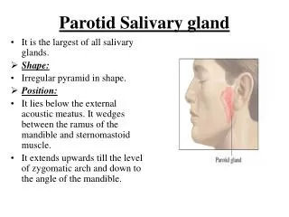
Parotid gland • 90% in superficial lobe, most in tail of gland. • Facial nerve is never paralyzed • “Dumb bell tumor” when arise from deep lobe.
Minor salivary gland: • lateral palate • submucosal mass
Histology • Tumor is typically well circumscribed • Some times may be uncapsulated or infltarated with tumor cells • Mixture of epithelial, myopeithelial and stromal components • Epithelial cells: nests, sheets, ducts, trabeculae • Stroma: myxoid, chrondroid, fibroid, osteoid • Tumor pseudopods
Diagnosis • History • Clinical examination • Investigation Biopsy CT scan or MRI
Treatment • The tumor is highly implantable, • Recurrence rate after primary surgery is about 5%. • If simple enucleation is performed , the recurrence rate is between 20-30%.
Warthin’s tumor • 6-8% of salivary gland tumors • Arise from striated duct cells. • 6-10% of parotid neoplasms • 3% with associated neoplasms
Clinical Presentation • Slow-growing, painless mass • Usually in the tail of parotid gland • Fluctuant , smooth soft and compressible • Cystic once contain mucoid fluid • Or some time solid in nature • Older Males • 10% bilateral
The tumor, at the right of the image, is well-demarcated from the adjacent parotid tissue and tends to shell out from it. Gross Pathology
histology • Composed of ductal epithelium & lymphoid stroma • Inner luminal layer is consist of tall columnar cells with centrally placed hyperchromatic nuclei • second layer is cuboidal cells • Stroma: • mature lymphoid follicles with germinal centers
Rating for S Gland tumors • Staging • (Treatment plan) • Grading • (Prognosis)
STAGING • TNM STAGING OF Salivary gland tumors
TNM STAGING • T = Tumor size • N = Lymph node involvement • M = metastasis
T Represents The Size Of The Primary Tumor • T0 - No primary tumor • Tis - Carcinoma in situ • T1 - Tumor 2 cm or smaller • T2 - Tumor 4 cm or smaller • T3 - Tumor larger than 4 cm without the involvement of Nerve • T4 - Tumor larger than 4 cm and deep invasion and nerve involvement
N - Represents Lymphatic Node Involvement • N0 - No nodes • N1 - Single homolateral node smaller than 3 cm • N2 - Nodes(s) homolateral smaller than 6 cm • N3 - Nodes(s) larger than 6 cm and/or bilateral
M- represents Tumor metastasis • M0 - No metastasis • M1 - Metastasis noted
Grading of Salivary gland tumors • High Grade • Low Grade • High/low grade • (Well, Moderate, Poor Differentiated)
Malignancy features • Usually tumor grows rapidly • Pain, Ulcerated, • Involvement of facial nerve • Involvement of skin • Involvement of L node • Metastasis: L,L,L • (Lung, Liver, Long Bone)
Mucoepidermoid Ca Major gland: • 90% arise in parotid, • 8% in the Submandibular, • 1% in the sublingual. Minor gland: more common: • 41% palate, • 14% buccal, • 9% tongue, • 5% lip.
Mucoepidermoid Ca • 4-9% of salivary gland tumors • 3rd-8th decades, • peak in 5th decade • Females more common • Most common salivary gland in children
Presentation • Low-grade: slow growing, painless mass • High-grade: rapidly enlarging, +/- pain, Ulceration
Gross pathology • Well-circumscribed to partially encapsulated to unencapsulated • Solid, • cystic, • mixed
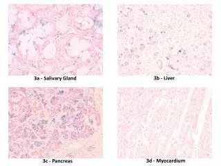
Mixture of mucous secrecting cells which are Cubodail or columnar in nature & squamous cells Histology
Adenoid Cystic Carcinoma • Overall 2nd most common malignancy • Most common in minor salivary gland, • Parotid gland, • Submandibular gland, • sublingual • M = F • 5th decade
Presentation • Asymptomatic enlarging mass • Ulcerated 50 % to 60 % • Pain, paresthesias, • Facial weakness/paralysis • Perinural invasion
Gross pathology Well-circumscribed Solid, rarely with cystic spaces Infiltrative
Tubular Pattern Solid Pattern Cribriform Pattern Histology