Download
1 / 35
360 likes | 368 Views
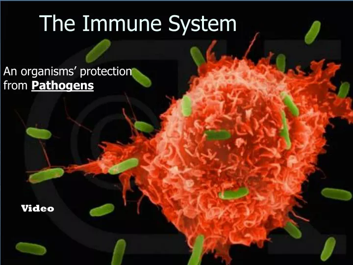
An organisms’ protection from Pathogens. The Immune System. Video. Pathogen. Bacteria Viruses Fungi Protists/parasites. Any infectious agent that causes disease. 2 Divisions of Immunity in Humans and Other Mammals. I. Innate Immunity – “Non-Specific”

E N D
An organisms’ protection from Pathogens The Immune System Video
Pathogen • Bacteria • Viruses • Fungi • Protists/parasites Any infectious agent that causes disease
2 Divisions of Immunity in Humans and Other Mammals I. Innate Immunity – “Non-Specific” This defense is not concerned with ‘what’ the pathogen is. This system merely prevents the pathogen from entering the body or destroys it before identifying it. It shoots first and asks questions later • Innate Immunity involves several layers of defense: • Barrier Defenses B. Inflammatory Response • C. Cellular Defenses D. Natural Killer Cells • E. Antimicrobial Defenses
A. Barrier Defenses • Epidermis – impenetrable barrier • Oil and Sweat have low pH • Resident Flora – your own bacteria • Mucous Membrane – mucus and cilia • Lysozyme
Skin • Epidermis an impenetrable barrier to pathogens • Oil and Sweat have a low pH that reduces pathogen growth • Resident Flora your own colonies of bacteria that live on your skin out-- competes with harmful bacteria for space
Mucous Membranes • Un-keritonized skin of the mocous membranes have a different layer of defense • MUCUS and CILIA Lysozyme
B. Cellular Defenses Microbes/antigens PHAGOCYTIC CELL Leukocytes – phagocytic white blood cells have surface receptors that detect typical pathogen compounds called antigens. Vacuole Lysosome containing enzymes
EXTRACELLULAR FLUID Groups of pathogens are recognized by TLR, Toll-like receptors Lipopolysaccharide Helper protein Flagellin TLR4 WHITE BLOOD CELL TLR5 VESICLE TLR9 CpG DNA Inflammatory responses TLR3 A white blood cell engulfs a microbe, then fuses with a lysosome to destroy the microbe
There are different types of phagocytic cells: • Neutrophils engulf and destroy microbes • Macrophages are part of the lymphatic system and are found throughout the body • Eosinophils discharge destructive enzymes • Dendritic cells stimulate development of acquired immunity
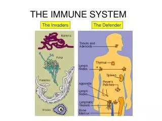
Fig. 43-7 Interstitial fluid Adenoid Adenoid Tonsil Tonsil Blood capillary Lymph nodes Blood capillary Lymph nodes Spleen Lymphatic vessel Tissue cells Spleen Lymphatic vessel Tissue cells Peyer’s patches (small intestine) Peyer’s patches (small intestine) Appendix Appendix Lymphatic vessels Lymph node Masses of defensive cells Lymph node Masses of defensive cells
C. Antimicrobial Proteins • Peptides and proteins function in innate defense by attacking microbes directly or impeding their reproduction • Interferon proteinsprovide innate defense against viruses and help activate macrophages • About 30 proteins make up the complement system, which causes lysis of invading cells and helps trigger inflammation
D. Inflammatory Responses • Following an injury, mast cells release histamine,which promotes changes in blood vessels; this is part of the inflammatory response • These changes increase local blood supply and allow more phagocytes and antimicrobial proteins to enter tissues • Pus, a fluid rich in white blood cells, dead microbes, and cell debris, accumulates at the site of inflammation
Fig. 43-8-3 D. Inflammatory Responses Pathogen Splinter Chemical Signals (Ligand) Macrophage Fluid Mast cell Capillary Phagocytosis Red blood cells Phagocytic cell
Fever is a systemic inflammatory response triggered by pyrogens released by macrophages, and toxins from pathogens • Septic shock is a life-threatening condition caused by an overwhelming inflammatory response
II. Acquired immunitylymphocyte receptors provide pathogen-specific recognition • Lymphocytes- are white blood cells that recognize and respond to antigens, foreign molecules. • Lymphocytes that mature in the thymus above the heart are called T cells,and those that mature in bone marrow are called B cells • Lymphocytes have immunological memory.
Fig. 43-9 B cells and T cells have receptor proteins that can recognize and bind to antigens Antigen- binding site Antigen- binding site Antigen- binding site Plasma membrane B cell Cytoplasm of B cell Cytoplasm of T cell T cell (a) B cell receptor (b) T cell receptor
Antibody Genes V V V D D J J J J C Heavy chain Light chain V D J C Antigen-binding region Constant region Assembled antibody molecule Rearranged gene components encoding a heavy chain Gene components scattered through one chromosome
Markers of Self: Major Histocompatibility Complex Antigenic peptide Antigenic peptide Antigenic peptide Viral infection MHC Class I MHC Class I MHC Class II Antigen-presenting cell uses MHC Class I or II Infected cell Cell membrane Body Cell with “Self-Markers called MHC
B Cells Antigen-specific B cell receptor Class II MHC and processed antigen are displayed Antigen Antibodies (Immunoglobins) B cell Cytokines (Lymphokines) LIGAND Plasma cell Activated helper T cell bacteria
T Cells Resting helper T cell Resting cytotoxic T cell Activated when they encounter infected cells that are presenting antigens Cytokines Released by Helper T-Cells Granule w/ destructive enzymes Activated helper T cell Activated killer cell
Killer Cells: Cytotoxic Ts Killer cell Target cell Target-oriented granules Surface contact
T cells bind to antigen fragments presented on a host cell • These antigen fragments are bound to cell-surface proteins called MHC molecules • MHC molecules are so named because they are encoded by a family of genes called the major histocompatibility complex
The Role of the MHC • In infected cells, MHC molecules bind and transport antigen fragments to the cell surface, a process called antigen presentation • A nearby T cell can then detect the antigen fragment displayed on the cell’s surface • Depending on their source, peptide antigens are handled by different classes of MHC molecules
Fig. 43-12 Microbe Antigen- presenting cell Infected cell Antigen associates with MHC molecule 1 Antigen fragment Antigen fragment 1 1 Class II MHC molecule Class I MHC molecule 2 2 T cell receptor T cell receptor 2 T cell recognizes combination (a) Cytotoxic T cell (b) Helper T cell
Activation of B Cells to Make Antibody Circulating antibody Antigen Antigen-presenting cell Antigen-specific B cell receptor Class II MHC and processed antigen are displayed Antigen is processed Antigen Class II MHC Cytokines (LIGAND) B cell Antibodies Antigen-presenting cell Activated helper T cell Plasma cell
Fig. 43-14 Antigen molecules B cells that differ in antigen specificity Antigen receptor Animation: Role of B Cells Antibody molecules Clone of memory B cells Clone of plasma cells
Fig. 43-16 Humoral (antibody-mediated) immune response Cell-mediated immune response Key Antigen (1st exposure) Stimulates Gives rise to + Engulfed by Antigen- presenting cell + + + B cell Helper T cell Cytotoxic T cell + + Memory Helper T cells + + + Antigen (2nd exposure) Memory Cytotoxic T cells Active Cytotoxic T cells + Plasma cells Memory B cells Secreted antibodies Defend against extracellular pathogens by binding to antigens, thereby neutralizing pathogens or making them better targets for phagocytes and complement proteins. Defend against intracellular pathogens and cancer by binding to and lysing the infected cells or cancer cells.
Fig. 43-17 Animation: Helper T Cells Antigen- presenting cell Peptide antigen Bacterium Class II MHC molecule CD4 TCR (T cell receptor) Cytokines Helper T cell + Humoral immunity (secretion of antibodies by plasma cells) + Cell-mediated immunity (attack on infected cells) + + B cell Cytokines Cytotoxic T cell
Cytotoxic T Cells: A Response to Infected Cells • Cytotoxic T cells are the effector cells in cell-mediated immune response • The activated cytotoxic T cell secretes proteins that destroy the infected target cell Animation: Cytotoxic T Cells
Fig. 43-18-3 Released cytotoxic T cell Cytotoxic T cell Perforin Granzymes perforin CD8 TCR granzymes Dying target cell Class I MHC molecule Pore Target cell Peptide antigen
B Cells: A Response to Extracellular Pathogens • The humoral response is characterized by secretion of antibodies by B cells • Activation of B cells is aided by cytokines and antigen binding to helper T cells • Clonal selection of B cells generates antibody-secreting plasma cells, the effector cells of humoral immunity
Fig. 43-19 Bacterium Antigen-presenting cell Peptide antigen B cell Class II MHC molecule Clone of plasma cells Secreted antibody molecules + Cytokines TCR CD4 Endoplasmic reticulum of plasma cell Activated helper T cell Helper T cell Clone of memory B cells 2 µm
The Role of Antibodies in Immunity • Neutralization occurs when a pathogen can no longer infect a host because it is bound to an antibody • Opsonization occurs when antibodies bound to antigens increase phagocytosis • Antibodies together with proteins of the complement system generate a membrane attack complex and cell lysis Animation: Antibodies
Fig. 43-21 Viral neutralization Opsonization Activation of complement system and pore formation Antibodies bound to antigens on viruses can neutralize the virus Bacterium Complement proteins Binding of antibodies to bacteria Promotes phagocytosis of the Bactria by Macrophages Virus Formation of membrane attack complex Flow of water and ions Macrophage Pore Foreign cell Following activation the attack complex forms pores in the foreign cell’s membrane , allowing water and ions to rush in. Binding of antibodies to antigens on the surface of a foreign cell activates a complex system. The Cell swells and eventually lyses.
Evolution and Immunity VIDEO 1 VIDEO 2