Download
1 / 29
290 likes | 300 Views
Lectures on Medical Bi o physics Department of Biophysics, Medical Faculty, Masaryk University in Brno. Structure of living matter. Water Properties of colloids Structure of proteins Structure of nucleic acids

E N D
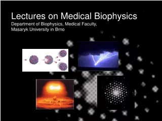
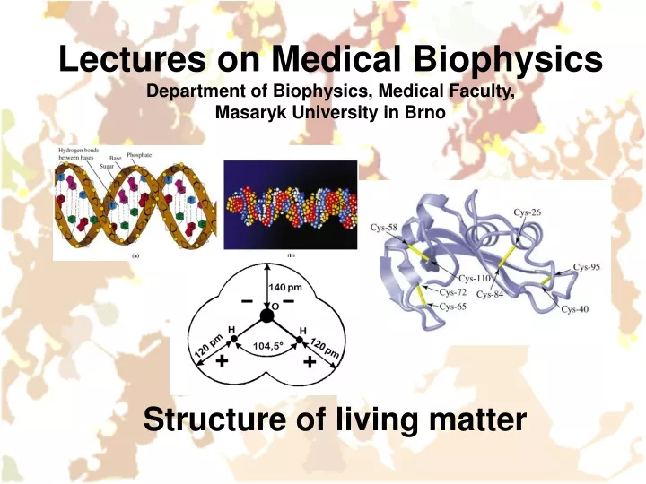
Lectures on Medical BiophysicsDepartment of Biophysics, Medical Faculty, Masaryk University in Brno Structure of living matter
Water Properties of colloids Structure of proteins Structure of nucleic acids This lecture deals only with selected components of living matter with distinct biophysical properties. Importance of some other components, e.g. electrolytes will be shown in the lecture on bioelectric phenomena. Check on further information in textbooks of biology and biochemistry. Lecture outline
Water Molecules of water are strongly polar. Moreover, between the oxygen and hydrogen atoms of neighbouring molecules, hydrogen bonds are formed. They join water molecules in aggregates – clusters.
Hydrogen bonds between water molecules Liquid water Ice Pictures: http://cwx.prenhall.com/bookbind/pubbooks/hillchem3/medialib/media_portfolio/11.html
Colloids – also known as non-true solutions – the solution consists of solute particles of diameter about 10 – 1000 nm dispersed in the solvent. We can distinguish two types of colloids according to the type of binding forces: Micellar colloids (also associative, small particles are bound together by van der Waals bonds) Molecular colloids (particles are macromolecules which subunits are bound together by covalent bonds) Colloids
Weak chemical bonds • Hydrogen bonds • Hydrophobic interaction • van der Waals bonds Also London forces, sometimes not classified as van der Waals bonds
Properties of colloids Mechanical: rigidity, elasticity, viscosity – caused by covalent and weak chemical bonds These properties depend on the form of colloid: sol (liquid) or gel (solid). Gel formation = gelatinisation Optical: • Light scatter: Tyndall effect (opalescence). Light can be scattered off the colloid particles. • Track of a light beam passing through a colloid is made visible by the light scattered by the colloidal particles. • Ultramicroscopy – before electron microscopy it was possible to observe colloidal particles in a light microscope as points of light on a dark background (observation in dark field). • Optical activity: Colloidal particles can rotate the plane of polarization of plane-polarised light passing through the colloid Electrical: see lecture on instrumental methods in molecular biophysics
Tyndall effect in micellar and molecular colloids - In solution of colloidal gold http://mrsec.wisc.edu/edetc/cineplex/gold/ - In solution of gelatin (a protein) http://link.springer-ny.com/link/service/journals/00897/papers/0006002/620095mb.htm
According to the affinity of the biopolymer to solvent (water) Lyophilic (hydrophilic) - form stable solutions Lyophobic (hydrophobic) - form unstable solutions According to the shape of the biopolymer (the shape is also influenced by the solvent!) Linear (fibrillar – DNA, myosin, synthetic polymers…..also scleroproteins, mostly insoluble in pure water) Spherical (globular – haemoglobin, glycogen … also spheroproteins, mostly soluble in pure water) Types of Colloids - Biopolymers
According to the products of hydrolysis: simple (only amino acids in hydrolysate) conjugated (not only amino acids in hydrolysate) Nucleoproteins Haemoproteins Flavoproteins Metalloproteins Lipoproteins ….. (see Biochemistry) Chemical composition of proteins
Structural units of proteins are amino acids (AA), connected by peptide bond: -RCH-NH-CO-RCH-, which can hydrolyse: -RCH-NH-CO-RCH- + H2O -RCH-NH2 + -RCH-COOH The carboxylic and amino groups can dissociate or protonise. E.g. the glutamic and asparagic acids have one free carboxylic group: -COOH -COO- + H+ AA lysine and arginine have one free amino group, which can protonise: -NH2 + H+ -NH3+ In proteins, 20 different AA can be found which can be divided into AA with polar and non-polar side chain. AA with aromatic ring or heterocycle (phenylalanine, tyrosine, tryptophan) strongly absorb UV light around 280 nm. AA cysteine contains sulphhydryl (thiol) group (-SH), which is oxidised by dehydrogenation and connected with dehydrogenated group of another cysteine residue by covalent disulphidic bridge (bond -S-S-). Structure of proteins
Structure of proteins Molar absorption coefficient e Disulphidic bridges stabilise the protein structure (bovine ribonuclease A) • http://cwx.prenhall.com/horton/medialib/media_portfolio/text_images/FG04_28a-b.JPG Wavelength (nm) Absorption spectrum of free phenylalanine, tyrosine and tryptophan in UV range • According:http://www.fst.rdg.ac.uk/courses/fs460/lecture6/lecture6.htm
Primary (sequence of covalently bound AA residues) Secondary (mutual spatial arrangement of neighbouring links of the polypeptide chain – given mainly by hydrogen bonds) a-helix b-structure (pleated sheet) other Tertiary (spatial arrangement of the polypeptide chain as a whole – given by hydrophobic and hydrogen bonds, stabilised by -S-S- bridges) Quaternary (a way of non-covalent association of individual polypeptide chains (subunits) in whole of higher order) Homogeneous – all subunits are identical Heterogeneous – subunits of two or more kinds Structure of proteins
Rise per residue • Podle: http://cwx.prenhall.com/horton/medialib/media_portfolio/text_images/FG04_10.JPG
b-structure (pleated sheet – antiparallel model)http://www-structure.llnl.gov/Xray/tutorial/protein_structure.htm
Triple helix of collagenhttp://cwx.prenhall.com/horton/medialib/media_portfolio/text_images/FG04_34.JPG
Podle: http://cwx.prenhall.com/horton/medialib/media_portfolio/text_images/FG04_01.JPG
Mononucleotide (the structural subunit of NA) is formed by: Pyrimidine (C, U, T) or purine (A, G) nitrogen base Sugar (ribose or deoxyribose) Phosphoric acid residue DNA: up to hundreds thousands of subunits. M.w. 107 – 1012. Two chains (strands) form antiparallel double helix. RNA: m-RNA (mediator, messenger) t-RNA (transfer) r-RNA (ribosomal) (viral RNA) Structure of nucleic acids (NA)
http://cwx.prenhall.com/horton/medialib/media_portfolio/text_images/FG19_13_90035.JPGhttp://cwx.prenhall.com/horton/medialib/media_portfolio/text_images/FG19_13_90035.JPG
B-DNAhttp://cwx.prenhall.com/horton/medialib/media_portfolio/text_images/FG19_15aC.JPGB-DNAhttp://cwx.prenhall.com/horton/medialib/media_portfolio/text_images/FG19_15aC.JPG
A-DNA – dehydrated, B-DNA – commonly present under physiological conditions, Z-DNA – in sequences rich on CG pairs
Superhelical structure of circular DNA • Podle http://cwx.prenhall.com/horton/medialib/media_portfolio/text_images/FG19_191C.JPG
Structure of chromatinhttp://cwx.prenhall.com/horton/medialib/media_portfolio/text_images/FG19_23_00742.JPG, http://cwx.prenhall.com/horton/medialib/media_portfolio/text_images/FG19_25_00744.JPG
Transfer RNA for valine – schematic • t-RNA from yeasts↓ • http://cwx.prenhall.com/bookbind/pubbooks/hillchem3/medialib/media_portfolio/text_images/CH23/FG23_14.JPG, http://www.imb-jena.de/cgi-bin/ImgLib.pl?CODE=4tra Amino acid binding site Valine
Ribosomal RNA • Next picture was published in: Science 11 February 2011: Vol. 331 no. 6018 pp. 730-736 in the article: Crystal Structure of the Eukaryotic 40S Ribosomal Subunit in Complex with Initiation Factor 1 (Julius Rabl, Marc Leibundgut, Sandro F. Ataide, Andrea Haag, Nenad Ban) Description: Architecture of the 40S. (A) Front and back views of the tertiary structure of the 40S showing the 18S rRNA as spheres and colored according to each domain (5′ domain, red; central domain, green; 3′ major domain, yellow; 3′ minor domain, blue; ESs, magenta), and the proteins as gray cartoons (abbreviations: H, head; Be, beak; N, neck; P, platform; Sh, shoulder; Bo, body; RF, right foot; LF, left foot). (B) Secondary structure diagram of the Tetrahymena thermophila(a protist)18S RNA …showing the rRNA domains and the locations of the ESs. (C) Ribosomal proteins of the 40S are shown as cartoons in individual colors; rRNA is shown as gray surface. The 40S is shown as in (A). (D) View of the quaternary interactions between ES6 and ES3 at the back of the 40S. The RNA is displayed as a cartoon with the proteins omitted for clarity. ES6 helices are colored in a gradient from light to dark magenta and labeled from A to E... ES3 is highlighted in pink, and the rest of the 18S rRNA is colored in gray. (E) The position of helix h16 in bacterial 30S [left…] and in 40S.
Changes in secondary, tertiary and quaternary structure of biopolymers are denoted as conformation changes. They can be both reversible and irreversible. ‘native’ state of a biopolymer: itsfunctional state. Otherwise the biopolymer has been ‘denatured’. Conformation changes and denaturation of biopolymers
Physical: Increased temperature Ionising radiation Ultrasound ….. Chemical: Changes of pH Changes in electrolyte concentration Heavy metals Denaturation agents destroying hydrogen bonds – urea ….. Combination of above factors: ionising radiation or ultrasound act directly and/or indirectly (chemically via free radicals) Denaturation factors
Author: Vojtěch MornsteinContent collaboration and language revision: Carmel J. Caruana, Viktor BrabecPresentation design: Lucie MornsteinováLast revision: September 2015