Download
1 / 77
800 likes | 873 Views
35. Sensors. Chapter 35 Sensors. Key Concepts 35.1 Sensory Systems Convert Stimuli into Action Potentials 35.2 Chemoreceptors Detect Specific Molecules or Ions 35.3 Mechanoreceptors Detect Physical Forces 35.4 Photoreceptors Detect Light. Chapter 35 Opening Question.

E N D
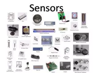
35 Sensors
Chapter 35 Sensors • Key Concepts • 35.1 Sensory Systems Convert Stimuli into Action Potentials • 35.2 Chemoreceptors Detect Specific Molecules or Ions • 35.3 Mechanoreceptors Detect Physical Forces • 35.4 Photoreceptors Detect Light
Chapter 35 Opening Question How do kangaroo rats, rattlesnakes, owls, bats, and moths “see in the dark?”
Concept 35.1 Sensory Systems Convert Stimuli into Action Potentials • Sensory receptor cells, or sensors or receptors, transduce physical and chemical stimuli into a change in membrane potential. • The change in membrane potential may generate an action potential that conveys the sensory information to the CNS for processing. • Sensory transduction—begins with a receptor protein that can detect a specific stimulus. • The receptor protein opens or closes ion channels in the membrane, changing the resting potential.
Concept 35.1 Sensory Systems Convert Stimuli into Action Potentials • Receptor potentials—graded membrane potentials that travel a short distance. • Receptor potentials can generate action potentials in two ways: • Can generate action potentials in the receptor cell • Can trigger release of neurotransmitter so that a postsynaptic neuron generates an action potential
Concept 35.1 Sensory Systems Convert Stimuli into Action Potentials • Stretch receptors in crayfish cause receptor potentials when the attached muscle is stretched. • Receptor potentials spread to the base of the axon and generate action potentials. • The rate of firing depends on the magnitude of the receptor potential, which depends on the amount of stretching.
Figure 35.1 Stimulating a Sensory Cell Produces a Receptor Potential
Concept 35.1 Sensory Systems Convert Stimuli into Action Potentials • Different sensory receptors respond to particular stimuli: • Mechanoreceptors detect physicalforces such as pressure (touch) and variations in pressure (sound waves). • Thermoreceptors respond to temperature. • Electrosensors are sensitive to changes in membrane potential.
Concept 35.1 Sensory Systems Convert Stimuli into Action Potentials • Chemoreceptors respond to the presence or absence of certain chemicals. • Photoreceptors detect light. • Some sensory receptor cells are organized with other cells in sensory organs, such as eyes and ears. • Sensory systems include sensory cells, associated structures, and neural networks that process the information.
Figure 35.2 Sensory Receptor Proteins Respond to Stimuli by Opening or Closing Ion Channels
Concept 35.1 Sensory Systems Convert Stimuli into Action Potentials • Sensation depends on which part of the CNS receives the sensory messages. • Intensity of sensation is coded as the frequency of action potentials. • Some sensory cells transmit information to the brain about internal conditions, without conscious sensation.
Concept 35.1 Sensory Systems Convert Stimuli into Action Potentials • Adaptation—diminishing response to repeated stimulation. • Enables animals to ignore background conditions but remain sensitive to changing or new stimuli. • Some sensory cells don’t adapt (e.g., mechanoreceptors for balance).
Concept 35.2 Chemoreceptors Detect Specific Molecules or Ions • Chemoreceptors—receptor proteins that bind to various molecules, responsible for taste and smell. • Also monitor internal environment, such as CO2 levels in blood. • Olfaction—sense of smell; depends on chemoreceptive neurons embedded in epithelial tissue at top of nasal cavity (in vertebrates).
Figure 35.3 Olfactory Receptors Communicate Directly with the Brain (Part 1)
Figure 35.3 Olfactory Receptors Communicate Directly with the Brain (Part 2)
Concept 35.2 Chemoreceptors Detect Specific Molecules or Ions • Axons from olfactory sensors extend to the olfactory bulb in the brain—dendrites end in olfactory hairs on the nasal epithelium. • Odorant—a molecule that activates an olfactory receptor protein • Odorants bind to receptor proteins on the olfactory cilia. • Olfactory receptor proteins are specific for particular odorants.
Concept 35.2 Chemoreceptors Detect Specific Molecules or Ions • When an odorant binds to a receptor protein, it activates a G protein, which activates a second messenger (cAMP). • The second messenger causes an influx of Na+ and depolarizes the olfactory neuron. • Many more odorants can be discriminated than there are olfactory receptors. • In the olfactory bulb, axons from neurons with the same receptors converge on glomeruli.
Concept 35.2 Chemoreceptors Detect Specific Molecules or Ions • Pheromones—chemical signals used by insects to attract mates. • Example: Female silkworm moth releases bombykol. Male has receptors for bombykol on the antennae. • One molecule of bombykol is enough to generate action potentials.
Concept 35.2 Chemoreceptors Detect Specific Molecules or Ions • Vomeronasal organ (VNO) is found in many vertebrates—specialized for pheromones • It is a paired tubular structure embedded in the nasal epithelium. • When animal sniffs, the VNO draws a sample of fluid over chemoreceptors in walls. • Information goes to an accessory olfactory bulb and on to other brain regions.
Concept 35.2 Chemoreceptors Detect Specific Molecules or Ions • Gustation is the sense of taste. • Taste buds—clusters of chemoreceptors. • Some fish have taste buds on the skin; the duck-billed platypus has taste buds on its bill. • Human taste buds are embedded in the tongue epithelium, on papillae. The sensory cells generate action potentials when they detect certain chemicals.
Figure 35.5 Taste Buds Are Clusters of Sensory Cells (Part 1)
Figure 35.5 Taste Buds Are Clusters of Sensory Cells (Part 2)
Concept 35.2 Chemoreceptors Detect Specific Molecules or Ions • Humans taste salty, sour, sweet, bitter, and umami—a savory, meaty taste. • “Salty” receptors respond to Na+ depolarizing the cell. • “Sour” receptors detect acidity as H+, and “sweet” receptors bind different sugars. • Umami receptors detect the presence of amino acids, as in MSG. • Bitterness is more complicated and involves at least 30 different receptors.
Concept 35.3 Mechanoreceptors Detect Physical Forces • Mechanoreceptors are cells that detect physical forces. • Distortion of their membrane causes ion channels to open and a receptor potential to occur. • This may lead to the release of a neurotransmitter.
Concept 35.3 Mechanoreceptors Detect Physical Forces • The skin has diverse mechanoreceptors: • Free nerve endings detect heat, cold, and pain. • Merkel’s discs: Adapt slowly, give continuous information. • Meissner’s corpuscles: Adapt quickly, give information about change.
Concept 35.3 Mechanoreceptors Detect Physical Forces • Ruffini endings: Deep, adapt slowly, react to vibrating stimuli of low frequencies. • Pacinian corpuscles: Deep, adapt rapidly, react to vibrating stimuli at high frequencies.
Concept 35.3 Mechanoreceptors Detect Physical Forces • Muscle spindles: Mechanoreceptors in muscle cells, called stretch receptors. • When muscle is stretched, action potentials are generated in neurons. • CNS adjusts strength of contraction to match load on muscle.
Concept 35.3 Mechanoreceptors Detect Physical Forces • Golgi tendon organ: Another mechanoreceptor, in tendons and ligaments. • Provides information about the force generated by muscle; prevents muscle tearing.
Concept 35.3 Mechanoreceptors Detect Physical Forces • Hair cells—mechanoreceptors in organs of hearing and equilibrium. • Hair cells have projections called stereocilia that bend in response to pressure. • Bending of stereocilia can depolarize or hyperpolarize the membrane.
Figure 35.8 Hair Cells Have Mechanosensors on Their Stereocilia (Part 1)
Figure 35.8 Hair Cells Have Mechanosensors on Their Stereocilia (Part 2)
Concept 35.3 Mechanoreceptors Detect Physical Forces • Auditory systems use hair cells to convert pressure waves to receptor potentials. • Outer ear: • Pinnae collect sound waves and direct them to the auditory canal. • The tympanic membrane covers the end of the auditory canal and vibrates in response to pressure waves.
Concept 35.3 Mechanoreceptors Detect Physical Forces • Middle ear—air filled cavity: • Open to the throat via the eustachian tube. Eustachian tubes equilibrate air pressure between the middle ear and the outside. • Ossicles—malleus, incus, stapes— transmit vibrations of tympanic membrane to the oval window.
Concept 35.3 Mechanoreceptors Detect Physical Forces • Innerear has two sets of canals—the vestibular system for balance and the cochlea for hearing. • The cochlea is a tapered and coiled chamber composed of three parallel canals separated by Reissner’s membrane and the basilar membrane.
Concept 35.3 Mechanoreceptors Detect Physical Forces • The organ of Corti sits on the basilar membrane—transduces pressure waves into action potentials. • Contains hair cells with stereocilia—tips are embedded in the tectorial membrane. • Hair cells bend and create a graded potential that can alter neurotransmitter release.
Concept 35.3 Mechanoreceptors Detect Physical Forces • Upper and lower canals of the cochlea are joined at distal end. • The round window is a flexible membrane at the end of the canal. • Traveling pressure waves of different frequencies will produce flexion of the basilar membrane.
Concept 35.3 Mechanoreceptors Detect Physical Forces • Different pitches, or frequency of vibration, flex the basilar membrane at different locations. • Action potentials stimulated by mechanoreceptors at different positions along the organ of Corti are transmitted to regions of the auditory cortex via the auditory nerve.
Concept 35.3 Mechanoreceptors Detect Physical Forces • Conduction deafness: Loss of function of tympanic membrane or ossicles. • Nerve deafness: Damage to inner ear or auditory nerve pathways. • Hair cells in the organ of Corti can be damaged by loud sounds. This damage is cumulative and irreversible.
Concept 35.3 Mechanoreceptors Detect Physical Forces • The vestibular system in the mammalian inner ear has three semicircular canals at angles to each other, and two chambers—the saccule and the utricle. • Hair cells sense position and orientation of head by shifting of endolymph. • Cupulae in canalscontain hair cell stereocilia—otoliths in membrane exert pressure and bend stereocilia.