Download
1 / 93
1k likes | 1.94k Views
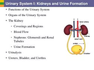
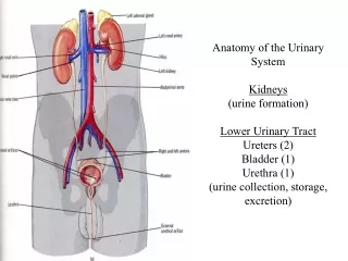
Urinary system Systema urinarium Kidneys Renes. Overview of urinary system. Upper urinary system Kidney ( Ren ) Nephron ( Nephron ) Collecting ducts (CD; Tubuli colligentes ) Lower urinary system Renal calices ( Calices renales ) Renal pelvis ( Pelvis renalis ) Ureter ( Ureter )

E N D
Overview of urinary system Upper urinary system • Kidney(Ren) • Nephron (Nephron) • Collecting ducts (CD; Tubuli colligentes) Lower urinary system • Renal calices (Calices renales) • Renal pelvis (Pelvis renalis) • Ureter (Ureter) • Urinary bladder (Vesica urinaria) • Urethra (Urethra)
Kidney = Ren • nephros in Greek • pl. nephroi • 150 g • facies anterior + posterior • extremitas superior + inferior • margo medialis + lateralis • hilum renale • sinus renalis • capsula fibrosa • lobi renales
Kidney – internal features • cortex: • labyrinthus • columnae renales protrude into medulla • medulla: • pyramides renales → papillae renales • area cribrosa + foramina papillaria • zona interna • zona externa • stria interna + externa • radii medullares protrude into cortex
Kidney microscopic structure • nephron • interstitium • vessels
Nephron • renal corpuscle • proximal tubule • intermediate tubule • distal tubule • juxtaglomerular apparatus
Renal corpuscleCorpusculum renale Malpighi • glomerulus (vascular convolut) • polus vascularis (vascular pole) • arteriola glomerularis afferens (wider) arteriola glomerularis efferens (narrower) • fenestrated (70-100nm) capillaries w/o diaphragma • capsula glomerularis (Bowmans pouch) • stratum parietale = parietal sheet • Flat single layer epithelium (epitheliocyti parietales) • stratum viscerale = visceral sheet • podocytes • spatium capsulare / urinarium • polus urinarius / tubularis (urinary pole)
Renal corpuscleCorpusculum renale Malpighi • podocytes • trabecules (cytotrabeculae) – primary extensions • pedicles (cytopediculi) – secondary extensions • basal membrane • mesangium (mesangium) • mesangial cells (mesangiocyti) • phagocyting, contractile and mitoting cells • mesangial matrix
Philtration barrier = barrier blood-urine (glomerular philter) 3 layers: • endothelium of glomerular capillaries • basal membrane • lamina rara interna + lamina densa (collagen IV, laminin, fibronectin, heparan sulphate) + lamina rara externa • pedicles of podocytes • interdigitate and form philtration cleft covered by cleft membrane – diaphragma rimae (nephrin) - other issues: size (10 nm) and substance charge → water permeable, low molecular substances → retain plasma proteins and blood cells
Mesangial cells (Mesangiocyti) • cells of mesenchymal origin • adjacent to capillary wall of glomerulus A: intraglomerular (= pericytes) • contractile function –receptors for angiotensin II and natriuretic factor (ANF) • supporting function • production of mesangial matrix and collagen • phagocytosis • proliferation B: extraglomerular mesangial cells • part of juxtaglomerular apparatus
Renal tubules 1. • proximal tubule (tubulus proximalis) • convoluted part (pars convoluta) • straight part (pars recta) • middle / intermediate tubule (tubulus intermedius) • descending limb (crus descendens) • ascending limb (crus ascendens) • distal tubule (tubulus distalis) • straight part (pars recta) • convoluted part (pars convoluta) • macula densa
Proximal tubule • simple cuboid epithelium • brush border on luminal side (limbus microvillosus) • striation on basal side = basolateral labyrinth (limbus plicatus basolateralis) – Na+-K+-ATPase • rich in mitochondria • resorption of NaCl and water (80-95%), glucose, aminoacids and proteins • Na+ into cell passively, from the cell activelly
Middle / intermediate tubule = thin part of Henle loop • flat cells, poor in organels • descending limb permeable for water • ascending limb not permeable for water • juxtamedullary nephrons have long Henle‘s loop • countercurrent system (together with vasa recta)
Henle‘s loopAnsa nephroni Henlei • formed by 3 morphological parts: • pars recta tubuli proximalis • tubulus intermedius • pars recta tubuli distalis • different length: • juxtamedullary nephrons have a long loop • cortical nephrons have a short loop • 5x more frequent compared to the long ones • parts: • thick descending limb • thin descending limb • thin ascending limb • thick ascending limb
Distal tubule • simple cuboid epithelium • cells are smaller compared to the proximal one • no brush border • striation on basal side = basolateral labyrinth (Na+-K+-ATPase) • rich in mitochondria • resorption of Na+ and secretion of K+ • regulated by aldosterone • macula densa • chemoreceptors (Cl- and Na+)
Juxtaglomerular apparatusComplexus juxtaglomerularis • granular cells of arteriola afferens + efferens = juxtaglomerular epitheloid cells (Juxtaglomerulocytus) • specialized smooth muscle layers of tunica media • mechanoreceptors • sympathetic innervation • production of renin • macula densa of distal tubule (epitheliocytus maculae densae) • approximately 30 slim cells • reverse polarita of cells • chemoreceptors • extraglomerular mesangial cells (mesangiocytus extraglomerularis Goormaghtighi; Lacis cell) • function: • blood pressure regulation - renin-angiotensin-aldosterone systém (RAA / RAAS)
Interstitium • connected with basal lamina of tubules and vessels • cortical x medullary • cell elements: • fibroblast-like cells • cells with adipous particle • macrophages, pericytes • non-cellular elements: • proteoglycans, glycoproteins, interstitial fluid
Renal tubules 2. • connecting tubule (tubulus reuniens) • oblouček mezi distálním a sběracím kanálkem • sběrací kanálek (tubulus colligens) • jednovrstevný kubický epitel • papilární vývod (ductus papillaris) • jednovrstevný cylindrický epitel • ústí na area cribrosa papillae renalis
Collecting ducts (Ductus colligentes) • principal cells (Epitheliocytus principalis) • concentration of urine • light cytoplasm, round nuclei • intercalated cells (Epitheliocytus intercalatus) • secretion of H+in exchange for K+ • reabsorption of bicarbonate(carboanhydrasis) • regulation of water resorption – antidiuretic hormone (adiuretin, vasopressin) = ADH • aquaporins
Kidney – arterial supply • a.renalis • paired visceral branch from aorta abdominalis at the level of discus intervertebralis L1/2, left one disc higher • a. renalis accessoria (30%) • caudally, branch from aorta abdominalis or a. iliaca communis / interna • blood flow 1,2-1,3 l/min • arteries are terminal = no arterio-arterial anastomoses →5 renal segments
Renal segments • segmentum superius • segmentum anterius superius • segmentum anterius inferius • segmentum inferius • segmentum posterius • individual vessel (r. posterior a. renalis)
Kidney – arterial supply2. a. renalis → r. anterior → 4 segmental branches → r. posterior for 1 posterior segment aa. segmentales → aa. lobares (approximately 12) → 2-3 aa. interlobulares → 2 vertically running aa. arcuatae → aa. interlobulares (aa. corticales radiatae) → arteriolae glomerulares afferentes → capillaries of glomerulus • blood pressure in glomerular system is higher (55 mmHg) → arteriolae glomerulares efferentes → peritubular capillary plexuses orarteriolae rectae along intermediate tubules of juxtaglomerular nephrons • blood pressure in the system of peritubular capillary plexus is lower (15 mmHg)
Kidney – venous drainage • vv. stellatae (from surface) • + venulae rectae (along intemediate tubules of juxtaglomerular nephrons) • + peritubular capillary plexus → → vv. interlobulares → vv. arcuatae → vv. interlobares → v. renalis → v. cava inferior • portal system(= rete mirabile) • 2 concomitant serially arranged capillary networks • strong veno-venous anastomoses
Kidney – lymph flow 3 lymph plexus peritubular, subcapsular and from capsula adiposa nodi lymphoidei lumbales plexus renalis autonomic, viscerosensory from ganglion coeliacum + plexus coeliacus from n. splanchnicus minor/imus via ganglion aorticorenale and plexus aorticus abdominalis Kidney – innervation
Resorptive function of kidneys countercurrent multiplication system different permeability of tubules for water and salts drainage of interstitium by vasa recta hormonal regulation RAA: renin →(from angiotensinogen in blood) angiotensin I (enzyme ACE on lung endotelium) angiotensin II (in suprarenal cortex)aldosterone ADH ANF other kidney hormones: erythropoetin, calcitriol